The Appendicular Skeleton
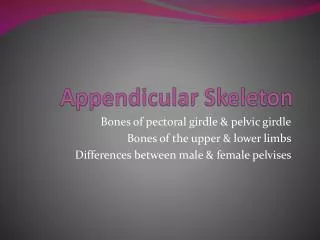
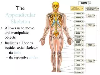
The Appendicular Skeleton. Composed of 126 bones Limbs (appendages) Pectoral girdle Pelvic girdle. The Appendicular Skeleton. acromion. Figure 5.6a. The Appendicular Skeleton. Figure 5.6b. The Pectoral (Shoulder) Girdle. Composed of two bones

The Appendicular Skeleton
E N D
Presentation Transcript
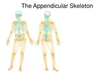
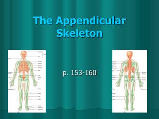
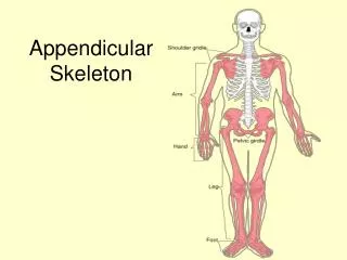
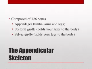
The Appendicular Skeleton • Composed of 126 bones • Limbs (appendages) • Pectoral girdle • Pelvic girdle
The Appendicular Skeleton acromion Figure 5.6a
The Appendicular Skeleton Figure 5.6b
The Pectoral (Shoulder) Girdle • Composed of two bones • Clavicle—collarbone –slender bone; at risk to fracture • Scapula—shoulder blade • These bones allow the upper limb to have exceptionally free movement • The clavicle serves as a brace to hold the arm away from the top of the thorax…so there is no problem with the arm clearing the wides dimension of the thoracic cage.
Bones of the Shoulder Girdle Coracoid process greater tubercle Figure 5.21a
Bones of the Shoulder Girdle CLAVICLE (#2) Figure 5.21b
Bones of the Shoulder Girdle * SCAPULA (#1) * * * Figure 5.21c–d
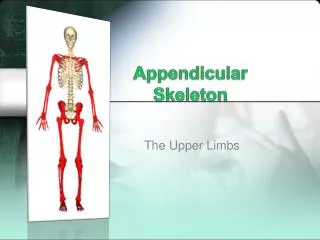
Bones of the Upper Limbs • Humerus • Forms the arm • Single bone
Bones of the Upper Limbs * Humerus (#3) * * * * * * * Figure 5.22a–b
Bones of the Upper Limbs • The forearm has two bones • Ulna • Medial bone in anatomical position • Radius • Lateral bone in anatomical position
Bones of the Upper Limbs Radius (#4) & Ulna (#5) * * * * Figure 5.22c
Bones of the Upper Limbs • The hand • Carpals—wrist 8bones • Metacarpals—palm • Phalanges—fingers 14 bones
Bones of the Upper Limbs Carpal (#6), Metacarpals (#7), Phalanges (#8) Figure 5.23
Bones of the Pelvic Girdle • Formed by two coxal (ossacoxae) bones • Composed of three pairs of fused bones • Ilium • Ischium • Pubis Pectoral Girdle is more flexible Pelvic Girdle is more secure and better able to bear weight
Bones of the Pelvic Girdle • The total weight of the upper body rests on the pelvis…therefore, must be massive • It protects several organs • Reproductive organs • Urinary bladder • Part of the large intestine • People instinctively curl over to protect internal organs
The Pelvis More massive than 4 legged creatures because it has to bear more weight. Figure 5.24a
Illium = yellow bone #1 Ischium = Purple bone #2 Pubis = Red bone #3 The Pelvis: Right Coxal Bone * * * * * * * * * * Figure 5.24b
Gender Differences of the Pelvis • The female inlet is larger and more circular • The female pelvis as a whole is shallower, and the bones are lighter and thinner • The female ilia flare more laterally • The female sacrum is shorter and less curved • The female ischial spines are shorter and farther apart; thus the outlet is larger • The female pubic arch is more rounded because the angle of the pubic arch is greater
Gender Differences of the Pelvis Figure 5.24c
True vs false pelvis • The greater or FALSE pelvis is located above the pelvic brim- SUPERIOR; supports the abdominal viscera the organs contained within the abdominal cavity; they include the stomach, intestines, liver, spleen, pancreas, and parts of the urinary and reproductive tracts • The lesser or TRUE pelvis below the brim- INFERIOR; limits delivery of baby
Bones of the Lower Limbs • The thigh has one bone • Femur • The heaviest, strongest bone in the body
Bones of the Lower Limbs Femur #4 * * * * * * Posterior view of right femur Anterior view of right femur Patella # 5 * * * * Figure 5.25a–b
Bones of the Lower Limbs • The lower leg has two bones • Tibia • Shinbone • Larger and medially oriented • Fibula • Thin and sticklike
Bones of the Lower Limbs * * Fibula #6 Tibia #7 * * * Figure 5.25c
Bones of the Lower Limbs • The foot • Tarsals • Two largest tarsals • Calcaneus (heelbone) • Talus • Metatarsals—sole • Phalanges—toes
Bones of the Lower Limb Talus #8 Calcaneus #9 Metatarsals #10 Phalanges #11 Figure 5.26
Arches of the Foot • Bones of the foot are arranged to form three strong arches • Two longitudinal • One transverse
Arches of the Foot Figure 5.27
Fallen Arches The ligament and tendons are weakend, allowing bones to “fall”