Download
1 / 48
770 likes | 2.34k Vues
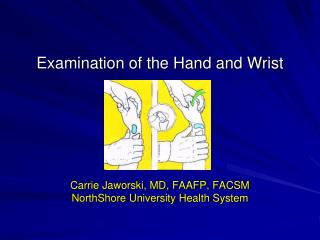
Examination of the Hand and Wrist. Carrie Jaworski, MD, FAAFP, FACSM NorthShore University Health System. Introduction. Superficial structures Easy palpation Must know anatomy Many structures, many diagnoses… Exam must correlate with imaging Contralateral hand/wrist as baseline.

E N D
Examination of the Hand and Wrist Carrie Jaworski, MD, FAAFP, FACSM NorthShore University Health System
Introduction • Superficial structures • Easy palpation • Must know anatomy • Many structures, many diagnoses… Exam must correlate with imaging • Contralateral hand/wrist as baseline
Anatomy Wrist • 8 bones: SLTPHCTT • Many Ligaments • Flexor and Extensor tendons • 3 nerves: • Ulnar • Median • Radial • Ulnar, radial arteries
Observation Position being held Ability to move, function Deformity Swelling Atrophy Color Scars
Wrist ROM Uninjured side as baseline Flexion 80 degrees Extension 70 degrees Ulnar deviation 30-35 degrees Radial deviation 20 degrees Pronation 75 degrees Supination 80 degrees
Strength Testing • Manual strength testing • 5 point scale • Resisted flexion, extension, ulnar and radial deviation, supination and pronation • Jamar dynamometer • 5 handle positions or one handle position with 3 measurements • Non-dominant hand as reference • Dominant hand generally 5-10% stronger • Women 60 lbs pressure, men 80-100 lbs pressure
Palpation Radial Wrist Dorsal Wrist Ulnar Wrist Palmar Wrist
Radial Wrist 1 • Distal palmar tuberosity of scaphoid • Ulnar/radial deviation • Flexor carpi radialis • First dorsal compartment • Radial styloid
Radial Wrist 2 • Dequervain’s • Finkelstein’s • Intersection syndrome • Junction of APL/EPB and ECRL/ECRB; 3-4 cm proximal to radial styloid • 1st CMC joint • Traction, ulnar pressure to reduce subluxation • Osteoarthritis • Grind test • Snuffbox • Ulnar deviation to palpate waist of scaphoid • Radial artery within
Dorsal Wrist Lunate Dorsal ganglion Extensor Digitorum (4th compartment) EDM (5th compartment) CMC joints
Ulnar wrist • TFCC • Grind test • ECU • Subluxation, painful snapping with supination of ulnarly deviated wrist • Ulnar styloid • Triquetreum • Radial deviation
Ulnar Wrist 2 • DRUJ • Piano key sign • Pisiform • Hook of hammate • FCU • Guyon’s canal
Palmar Wrist Palmaris Longus Carpal Tunnel injection
Special Tests Finklestein’s Tinel’s Phalen’s
Scapholunate Instability • Watson’s test • Thumb on palmar distal tuberosity • Ulnar to radial deviation • Scaphoid should move palmarly under thumb • Release with thumb, clunk suggests injury • Finger Extension test • Wrist in flexion • Resist finger extension over DIP joints • Pain SL interval suggests injury
Lunotriquetral Instability • Compression test • Thumb applies pressure radially to triquetrum between FCU and ECU • Ballottement test(Shuck) • Support lunate with thumb dorsally and index palmarly • Alternating dorsal and palmar loading of triquetrum with thumb and index of other hand
Radiocarpal Instability • Drawer • Support forearm with one hand • Grasp metacarpals with other, gentle traction, then anterior/posterior force • Alternative to grasp radius, then anterior/posterior force over triquetrum
Nerve/Vascular Impingement • Median Nerve • Sensation digits 1-4 • Thenar atrophy • Phalen’s • Tinel’s • Ulnar Nerve • Sensation digits 4-5 • Hypothenar atrophy • Ulnar artery thrombosis • Allen test
Anatomy-Hand • Palmar Creases • Distal Palmar Crease • Proximal Palmar Crease • Thenar Crease • Dorsal surface • MCP and IP joints • Nails
Anatomy-Hand • Bones • Metacarpals • 2nd and 3rd are immobile • 4th and 5th are mobile • 14 phalanges
Anatomy-Hand • Muscles (intrinsic) • Thenar and hypothenar • Pinching • Interossei • Lumbricals • 4 muscles that arise from the tendon of flexor digitorum profundus muscle. • IP extension • MCP flexion
Anatomy-Hand • Ulnar Nerve • Passes through Guyon’s canal with ulnar artery • Motor • Innervates all intrinsics except thenar muscles and 2 radial lumbricals • Power Grip • Sensory • Ulnar 1.5 • Testing • Finger abduction against resistance • Purest sensory test is palmar surface of tip of 5th finger
Anatomy-Hand • Median Nerve • Passes through carpal tunnel on volar wrist • Motor • Fine control of pincer grasp • Innervates thenar muscles and 2 radial lumbricals • Sensory • Radial 3.5 fingers and their dorsal tips • Testing • Opposition of thumb to each finger • Purest sensory test is palmar tip of index finger • Anterior Interosseus injury if can’t make “ok” sign
Anatomy-Hand • Radial Nerve • Motor • Innervates extrinsic wrist and finger extensors • Does not innervate any intrinsic muscles • Sensory • Dorsally for 3.5 fingers • Testing • Wrist and hand extension against resistance • Purest sensory test is web space between thumb and index fingers
Anatomy-Hand • Extensor tendons • Flexor tendons • FDP splits FDS to attach at distal phalanx • Test FDP by stabilizing PIP and flex DIP • Test FDS by anchoring other fingers in extension
Anatomy-Hand Blood supply: • Neurovascular bundles • Contain digital artery, vein, and nerve • Two bundles: one radial and the other ulnar • Radial and Ulnar arteries join in 2 arches • Superficial Palmar Arch: superficial to flexor tendon • Located at base of first web space • Deep Palmar Arch (deep to flexor tendons) • Proximal to superficial arch by 1 cm
Hand ROM • Abduction/Adduction – Abd 20 degrees • Thumb opposition • Thumb palmar abduction/adduction = 70/0 • Finger flexion/extension • MCP = 90/30-45 • PIP = 100/0 • DIP = 80-90/0-10 • Thumb IP = 80-90/0-20 • Thumb MCP = 55/0
Strength testing-hand • Grip • Pincer • Abduction • Adduction
Palpation • Thenar and hypothenar muscles • First Metacarpal • Metacarpals 2-5 • Phalanges • Collateral stability
Hand Injuries/Conditions • Osteoarthritis • Heberden’s nodes • Bouchard’s nodes
Central Slip Extensor Tendon Injury • Tender at dorsal aspect of the PIP joint (middle phalanx) • Inability to actively extend thePIP joint • Splint in full extension for 6 weeks • Refer: Avulsion fracture involving more than 30 percent of the joint or inability to achieve full passive extension
Boutonniere Deformity • Can occur acutely, but more often after several weeks • Extensor tendon/Central slip ruptures at PIP • Lateral bands slip volar and flex PIP, DIP extends
Extensor tendon injury-Mallet finger • Tear or stretch of extensor tendon prior to insertion on distal phalanx • Exam: Soft tissue swelling, lack of full extension of DIPJ
Mallet finger • X-ray may show lack of full extension with or without a fracture of proximal aspect of distal phalanx • Strict immobilization in full extension 6-8 weeks • Consider surgery for fx > 30% of articular surface
Flexor tendon injury-Jersey finger • Inability to actively flex distal phalanx • Ring finger most commonly affected • Protrudes further than other fingers on grasping • Forced extension of actively flexed DIP joint • Examples • Football player grabs a player's jersey on tackle • Lifting latch on car door
Jersey Finger • Avulsion of Flexor Digitorum Profundus (FDP) as DIP is forcibly extended • Can be seen with a laceration of the volar aspect of the phalanx • Tendon may retract to the PIP or as far as the palm • Surgical referral
Collateral Ligament Injury • Maximal tenderness at involved collateral ligament • Test stability of joint while the finger is in 30 degrees of flexion and the MCP joint is flexed. • Stable joint: buddy tape for two to four weeks. Do not leave fifth digit exposed if ring finger is taped.
Volar Plate Injury • Maximal tenderness at the volar aspect of involved joint • Test for full flexion and extension as well as collateral ligament stability. • Splint at 30 degrees of flexion and progressively increase extension for two to four weeks.Buddy tape at the joint if injury is less severe. • Refer: Unstable joints or large avulsion fragments
UCL Injury-Skier’s Thumb • AKA “gamekeeper’s thumb” • Caused by hyperextension of Ulnar collateral ligament • Exam: • Tender at UCL = x-ray first • Abduction stress at MCP with MCP in flexion • Abnormal if > 15 degrees from opposite side, or 35 degrees absolute
Skier’s thumb • Stenar lesion = surgery • Stable injuries are splinted • Can also get radial collateral ligament injuries
Metacarpal neck fractures • Check angulation and rotation • Angulation of 2nd, 3rd, 4th, 5th MC • Think: 10, 20, 30, 40 • Accept: 10,10, 30, 40 • Powergrip of index and long compromised by angulation therefore reduce anything >10 degrees