Download
1 / 13

E N D
X-radiation X-rays are part of the electromagnetic spectrum. X-radiation (composed of X-rays) is a form of electromagnetic radiation. X-rays have a wavelength in the range of 10 to 0.01 nanometers, corresponding to frequencies in the range 3 × 10¹⁶ Hz to 3 × 10¹⁹ Hz and energies in the range 120 eV to 120 keV. They are shorter in wavelength than UV rays and longer than gamma rays. In many languages, X-radiation is called Röntgen radiation, after Wilhelm Conrad Röntgen, who is generally credited as their discoverer. X-rays from about 0.12 to 12 keV (10 to 0.10 nm wavelength) are classified as "soft" X-rays, and from about 12 to 120 keV (0.10 to 0.010 nm wavelength) as "hard" X-rays, due to their penetrating abilities. X-rays are a form of ionizing radiation, and exposure to them can be a health hazard.
Bremsstrahlung and characteristic X-rays • There are two different atomic processes that can produce X-ray photons. One is called Bremsstrahlung and is a German term meaning "braking radiation“. • The other is called characteristic X-rays(K-shell emission). They can both occur in the heavy atoms of tungsten. Tungsten is often the material chosen for the target or anode of the x-ray tube.
BremsstrahlungX-rays • Electrons are scattered elastically and inelastically by the positively charged nucleus. The inelastically scattered electron loses energy, which appears as bremsstrahlung. Elastically scattered electrons (which include backscattered electrons) are generally scattered through larger angles than are inelastically scattered electrons.
Characteristic X-rays • An incident electron ionizes the sample atom by ejecting an electron from an inner-shell (the K shell, in this case). De-excitation, in turn, produces characteristic X-radiation (above) or an Auger electron (below). Secondary electrons are ejected with low energy from outer loosely bound electron shells, a process not shown.
Characteristic X-rays • When outer-shell electrons drop into inner shells, they emit a quantized photon "characteristic" of the element. The energies of the characteristic X-rays produced are only very weakly dependent on the chemical structure in which the atom is bound, indicating that the non-bonding shells of atoms are the X-ray source. The resulting characteristic spectrum is superimposed on the continuum. An atom remains ionized for a very short time (about 10-14 second) and thus the incident electrons that arrive about every 10-12 second can repeatedly ionize an atom. However, not all outer-shell electrons can fall in to produce X-rays.
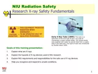
Coolidge side-window tube (scheme) • K: filament (-) • A: anode (+) • Win and Wout: water inlet and outlet of the cooling device (C) X-rays are generated by an X-ray tube. It works with a very good quality vacuum. The electrons are produced by thermionic effect from a metal filament heated by an electric current. The filament is the cathode of the tube. The high voltage potential is between the cathode and the anode, the electrons are thus acceleratedand then hit the metal target anode. X-ray tube.
The voltages used in diagnostic X-ray tubes, and thus the highest energies of the X-rays, range from roughly 20 to 150 kV. The anode is usually made out of tungsten or molybdenum. The high energy electrons interact with the atoms in the anode. Two atomic processes of production of x-ray photons. One is called Bremsstrahlung, which is a fancy German name meaning "braking radiation." The electron (much lighter than the nucleus) comes very close to the nucleus and the electromagnetic interaction causes a deviation of the trajectory where the electron looses energy and an X-ray photon is emitted. High energy electron beam striking high-Z material produces X-rays and Heat. 99% or more of the electron beam energy is deposited as heat! Less than 1% of electron beam energy is converted in X-rays! The ideal situation would be if most of the electrons created x-ray photons rather than heat. The energy of the emitted photon can take any value up to a maximum corresponding to the energy of the incident electron. The highest X-ray energy is equal to an electron kinetic energy:
The total flux of X-radiation is: I - tube current U – anode voltage Z – atomic number The anode has two primary functions: (1) to convert electronic energy into x-radiation, and (2) to dissipate the heat created in the process. The material for the anode is selected to enhance these functions. Another atomic process is X-ray fluorescence. If the electron has enough energy it can knock an orbital electron out of the inner electron shell of a metal anode atom, and as a result electrons from higher energy levels then fill up the vacancy and X-ray photons are emitted. This process produces an emission spectrum of X-ray frequencies, sometimes referred to as the spectral lines. The spectral lines generated depend on the target (anode) element used and thus are called characteristic lines. Usually these are transitions from upper shells into K shell (called K lines), into L shell (called L lines) and so on.