Download
1 / 20
200 likes | 361 Vues
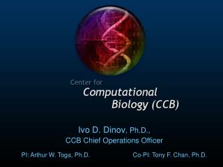
Ivo D. Dinov , Ph.D., CCB Chief Operations Officer PI: Arthur W. Toga, Ph.D. Co-PI: Tony F. Chan, Ph.D. AWT. CCB Overall Organization. Core 1: Computational Science Registration Shape Modeling Surface Modeling Segmentation. Core 2: Computational Tools Analysis Data Integration

E N D
Ivo D. Dinov, Ph.D., CCB Chief Operations Officer PI: Arthur W. Toga, Ph.D. Co-PI: Tony F. Chan, Ph.D. AWT
CCB Overall Organization Core 1: Computational Science Registration Shape Modeling Surface Modeling Segmentation Core 2: Computational Tools Analysis Data Integration Knowledge Management Core 3: Driving Biological Projects Brain Development Aging & Dementia Multiple Sclerosis Schizophrenia Core 4: Infrastructure/Resources Computing Software Informatics Core 5: Education & Training Courses Fellowships Workshops Training Materials Core 6: Dissemination Web Publications Education Database Core 7: Administration & Management Committees, SIGs Science Advisory Board Meetings & Communication Progress & Monitoring Support
CCB Major Objectives • Establish a new integrated multidisciplinary researchcenter in computational neurobiology. • Develop Atlases – sets of maps on different spheres of biological information that span many resolution-scales, image-modalities, species, genotypes & phenotypes. • Introduce new mathematical symbolic representations of biological information across space & time. • Develop, implement and test computational tools that are applicable across different biological systems & atlases.
CCB Grand Challenges • Brain Mapping Challenges • Software & Hardware Engineering Challenges • Infrastructure & Communication Challenges • Data Management • Multidisciplinary Science Environment
CCB Brain Mapping Challenges • Quantitative analysis of structural & functional data • Merging NeuroImaging and Clinical data (e.g., NPI) • NeuroImaging markers associated with Gender, Race, Disease, Age, Socioeconomics, Drug effects • NeuroImaging Interactions w/ Genotype-Phenotype • Understanding Temporal Changes in the Brain • Data Management (volume, complexity, sharing, HIPAA) • NeuroImaging Across Species (similarities and diff) • Integrating Multimodal Brain Imaging Data • Efficient and Robust Neurocomputation (Grid) • SW & Tool Development and Management (Pipeline)
Core 1 Specific Aims • Non-Affine Volumetric Registration • Parametric & Implicit Modeling of Shape & Shape Analysis using Integral Invariants • Conformal Mapping (on D2 or S2) • Volumetric Image Segmentation
Core 2: Computational ToolsResearch Categories • Data Analysis • Volumetric segmentation • Surface analyses • DTI Analysis (tractography) • Biosequence analysis • Interaction • Grid Pipeline Environment • SCIRun/Pipeline integration • New tools for integrating, managing, modeling, and visualizing data • Knowledge Management – Analytic strategy validation
Data Visualization Mutation Pathways Of HIV-1 Protease Additional functionality Is integrated via the extension architecture.
CCB – Driving Biological Projects (current) DBP 1: Mapping Language Development Longitudinally DBP2: Mapping Structural and Functional Changes in Aging and Dementia DBP3: Multiple Sclerosis and Experimental Autoimmune Encephalomyelitis DBP 4: Correlating Neuroimaging, Phenotype and Genotype in Schizophrenia
CCB – Driving Biological Projects (pending!) DBP 5 (Jack van Horn, Dartmouth): Computational Mining Methods on fMRI Datasets of Cognitive Function DBP 6 (Srinka Ghosh & Tom Gingeras, Affymetrix): Maps of Transcription and Regulation of key Brain tissuesin the Human Genome DBP 7 (James Gee, U Penn): Shape Optimizing Diffeomorphisms for Atlas Creation DBP 8 (Wojciech Makalowski, Penn State U): Alternative Splicing of Minor Classes of Eukaryotic Introns
The continuing CCB developments since publication include: New algorithms for brain surface representation, cortical thickness and variation Updates on efficiency of conformal mapping techniques Better synergy of multi-disciplinary resources *Genus Zero Surface Conformal Mapping and Its Application to Brain Surface Mapping Xianfeng Gu, Yalin Wang, Tony F. Chan, Paul M. Thompson and Shing-Tung Yau IEEE Transactions on Medical Imaging, 2004, Volume 23, Number 8 Modeling - Brain Conformal Mapping Last year’s groundbreaking publication* on conformal mapping as applied to brain surfaces initiated a novel technique for examining neuroscience data.
CCB Neuroimaging Applications:Brain Mapping of Disease CCB as featured in US News & World Report, 3/21/05 http://www.usnews.com/usnews/health/articles/050321/21brain.htm Mapping Schizophrenia Mapping temporal structural changes Schizophrenia. The disease causes a mix of hallucinations and psychotic behavior in teenagers. Abnormalities in schizophrenics first cropped up in the parietal lobe. Drug effects on the Brain Differences of antipsychotic drug effects. Alzheimer’s Disease Mapping Temporal anatomical alterations in Alzheimer’s disease. Gray matter loss starts in the hippocampus, a memory area, and quickly moves to the limbic system, which is involved in emotions.
Neuroimaging Applications:Beyond the Brain into the Mind CCB as featured in National Geographic magazine, Mach 2005 http://magma.nationalgeographic.com/ngm/0503/feature1/index.html CCB reaches 5 million readers via National Geographic and shares neuroscience research with the public. The CCB receives many requests from doctors and teachers interested in using these models as teaching devices. CCB’s 3D models show fMRI activity in the visual system, fear, meditation, navigation, musical pitch, object permanence, plasticity, autism and hypergraphia.
CCB Infrastructure (Core 4) SA-1: Computing Infrastructure Develop, implement and maintain the computing resources and network services required for computationally intensive science performed in the CCB SA-2: Application Deployment Integrate the algorithms, techniques and tools developed in Cores 1 & 2 with the Computing Infrastructure to enable researchers to remotely access and use the computing resources of the CCB SA-3: Computational Research Support Provide technical support and expertise to enable collaborators to use the resources of the CCB
CCB Education & Training (Core 5) • Coursework in imaging-based Computational Biology • Graduate & undergrad training in Computational Biology • Fellowship Program • Visiting Scholars Program • Workshops, Retreats & Tutorials • Educational Materials
Develop level set reps. for open curves/surfaces Test Cost functions for 2D Matching Test Cost functions for 3D Matching Ensure deformation mappings are diffeomorphic Test on 2D brain data Test on 3D brain data Add intensity information (Jensen divergence) Formal Validation in 2D and 3D Use by DBPs and the rest of the world Year 1 (10/04-3/05) Year 1.5 (4/05-9/05) Year 2 (10/05-3/06) Year 2.5 (4/06-9/06) Year 3 (10/06-3/07) Year 3.5 (4/07-9/07) Year 4 (10/07-3/08) Year 4.5 (4/08-9/08) Year 5 (10/08-3/09) Year 5.5 (4/09-9/09) Develop Multi-Layer Level Sets for 2D MRI Extend Multi-Layer Level Sets to Volumetric Data (3D) Extend Multi-Layer Level Sets to measurement of cortical thickness - Local modification of Image forces to improve overall segmentation Extend to topology preserving multi-layer level sets Validate on pediatric and adult data sets Develop Logic Models using Level Sets Apply to multi-channel segmentation of different modalities of MRI data Extend to un-registered images Detect pathology in one of the modalities Collect appropriate multi-sequence data sets and validate algorithms Perform a detailed analysis of the sensitivity of the algorithm to initial parameter selection Improve auto-matic initialization Extend algorithm from 2D to true 3D Local modification of the image forces to improve segmentation accuracy Integrate with multi-layer Level Sets to enable segmentation of cortical thickness Validate the algorithm across diverse data sets Extend 2-D Charged Fluid simulation to 3-D Apply 3-D Charged Fluid to volumetric 3-D image segmentation Apply the 3-D Charged Fluid to volumetric 3-D vascular image segmentation MR, CT and 3DRA) Adapt the particle size to object and boundary characteristics Validate the algorithm across diverse data sets (MR, CT and 3DRA) 5-1 6-1 6-2 6-3 6-4 Experimental evaluation of limitations of local and global shape representations Shape matching based on local descriptors Shape matching based on global deformations Kernel shape statistics with local priors Shape representation: hierarchy and compositionality.Convergence of local/global representations 3-D Shape descriptors and integral invariants 3-D Shape matching Dynamic shape signatures Classification of dynamic shapes Integration with other Cores Hippocampal Morphometry Studied with Brain Conformal Mapping Matching Landmarks 3D Paint Foliation and conformal Maps Shape Space Image Manifold Solving PDE on Surfaces with Conformal Structure Timeline for Core 1: Computational Science SA 1-2: Modeling of Shape and Shape Analysis SA: 1-1: Registration using Level Sets SA: 1-3:Parametric & Implicit Surface Models SA: 1-4:Volumetric Image Segmentation
Timeline for Core 2 : Computational Tools SA 1-2: Modeling of Shape and Shape Analysis Extend tissue classification methods to process multiple modalities and identify pathologic structures Image Segmentation Apply level set methods from Core I, Aim 4 to identify structures in MRI Combine level-set segmentation methods with atlas-based approaches to label neuro-anatomical structures. Extend methods to other modalities and specimens (mice) Validation – ongoing through duration of the project Develop methods for parameterizing zero-genus surfaces P-harmonic method validation Surface methods Application of parameterization from Core I Develop novel approaches for labeling cortical landmarks Develop tools for computing various measures from DTI data. DTI analysis Develop a fluid-model approach to fiber tract segmentation in DTI. Develop a DTI phantom model for validation ofDTI analysis algorithms Biosequence analysis Clinical Applications : Concurrent fMRI / DTI in surgical planning of tumor patientsMS Alzheimer's EAE models (mouse) Bio-sequence atlas tools Develop analysis tools and database technology for analyzing the role of alternative splicing in temporal development of neuronal tissue and disease states. CCB Pipeline Processing Environment SQL integration Tools OntologyNeuroimaging Domain Ontology Extension architecture implementation Grid engine integration Pipeline V.3 Public release Pipeline V.4 Public release Pipeline V.5 Public release BIRN SRB integration Networking API Library SCIRun integration Provenance integration Natural language interface Overlay networkgrid computing Compile CCB pipeline with SCIRun2 component model interface for LONI modules Connect LONI to SCIRun Connect LONI to ITK Enhanced user interface SCIRun Integration Shiva Brain GraphBAMS interface Integration withPipeline Surface Models Image Processing and Visualization PluginsDev support, API help, End user documentation LONI viz Redesign LONI_Viz core - small/tight core plus a diverse & expandable plug-in infrastructure Tool integration - LONI_Viz, SHIVA, Pipeline, SCIRun, Slicer Database Tools Biospeak for Comp. BiologyBLASTgres extension BioPostgres 1D/2D/3D atlasConDuit Useful container lib CompAtlas Computational Atlas Kernel