Download
1 / 41
440 likes | 668 Vues
Electrophysiology of Photoreceptors (from counting photons in starlight to the blazing sun snowy slopes). Phototransduction Cascade quick review Single Cell responses Currents, voltages transmitter release Rod and cone response differences. RPE Cells Photoreceptors Müller Cells. Outer

E N D
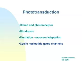
Electrophysiology of Photoreceptors (from counting photons in starlight to the blazing sun snowy slopes) Phototransduction Cascade quick review Single Cell responses Currents, voltages transmitter release Rod and cone response differences
Outer Segments Ellipsoid Synaptic region Salamander Rod and Cone cell Brett Gerwin MSII
Circulating current between the OS and IS in the dark partially depolarizes the cells. Light triggers HYPERPOLARIZATION and decreased transmitter release. Glutamate is the neurotransmitter. Biochemical cascade initiated by absorption of one photon by chromophore (11-cis retinal). Activated opsin acts as an enzyme. Rhodopsin and cone opsins are the classical G-protein couple receptor (GPCR). Opsin activates transducin, which activates phosphodiesterase (PDE). Activated PDE destroys cGMP cGMP is the 2nd messenger that keeps cation channels open Rods AND Cones
Detecting A Single Quantum In rods a single photon generates an electrical signal that is sufficiently above the noise so as to be reliably detectable.
Photocurrents are graded responses to lightthat changes membrane voltage which in turn drives neurotransmitter release
Rod sensitivity is high at a cost of speed, slow temporal sensitivity Toad rod recording at 20°C
Rod shaped OS Separate discs Slower pigment regeneration (renewal) Synaptic ending is small round spherule with few ribbons Connect only to On-type, rod bipolars Cone shaped OS Fused discs, continuous with extracellular space Pedicle shaped synaptic terminal with More ribbon synapses (20) Connects to many types of BOTH on & off Bipolar cells Rods vs. Cones
Very stable visual pigment Greater biochem gain Slower responses Lower Ca++ permeability through cGMP channel Saturation Limited operating range Less stable visual pigment Lower sensitivity(gain) Faster temporal response Greater Ca++ permeability through cGMP channel CONES NEVER SATURATE to steady light. 10x faster PDE inactivation. Rods vs. Cones Biochemical
A look at rod responses: the #’s Chipmunk Rod: I1/2 = 200 photonµm -2 Sf = 0.090 pA-photon -1µm2 ttpeak = 100 msec Ti =90 msec
Cone responses: the number’s Chipmunk Cones: I1/2 = 7,000 photon- µm-2 Sf =0.0005 pA-photon-1µm2 ttpeak = 50 msec Ti = 40 msec
ROD/CONE Ratios Chipmunk Rod: I1/2 = 200 photonµm -2 Sf = 0.090 pA-photon -1µm2 ttpeak = 100 ms Ti =90 ms Chipmunk Cones: I1/2 = 7,000 photon- µm-2 Sf =0.0005 pA-photon-1µm2 ttpeak = 50 msec Ti = 40 ms Rod/Cones: I1/2 : 1/35 Sf : 1/180 ttpeak : 2 Ti : 2.25
Inactivation steps control sensitivity and timing • ROD and CONE transduction are different! • 2x timing • 50 to 100x in sensitivity • Although the specific details of the differences is not yet known. . . . • Kinases for phosphorylation of R* differ • The cGMP gated channels are different • GCAP proteins that are Ca++ sensitive feedback signal are different • Inactivation of PDE* by RGS9 are probably different (Cones have 10x rod levels of RGS9)
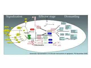
Phototranscduction Cascade • Photon of light generates R* • Stage 1: R* collides with G protein (both on the membrane disk) (500 to 800 fold amplification) • Gabg-GDP : R* promotes exchange of GTP for GDP • G protein splits to become active Ga-GTP and the Gbg • Stage 2: Ga-GTP collides with and attaches to the enzyme PDE (gabg) complex dislodging an inhibitory unit (PDEg) Gain factor 1. • The Ga-GTP- PDE complex greatly enhances PDE activity • Stage 3: activated PDE hydrolyzes cGMP -> 5’ GMP • Gain factor 6-50 • TOTAL GAIN about 5000 cGMP destroyed, 1,000,000 Na/Ca ions excluded from outer segment of rod photoreceptor outer segment.
Cyclic activity of enzymes in Cascade RGS9/G5/R9AP
Increasing RGS9/G5/R9AP proteins 25 fold alters the rod response.
Calcium Feedback Light closes the outer segment cation channel reducing influx of Ca2+, a potent feedback signal in phototransduction.
Calcium Feedback • Guanylate cyclase replaces the cGMP to reopen the channel to repolarize the membrane back to resting levels. • Cyclase activity is cubically dependent on Ca2+ • Calmodulin is a calcium binding protein that interacts with the cGMP channel to modulate cGMP binding affinity. • Recoverin is modulated by Ca2+ and is part of the rhodopsin recovery pathway.
Shutting off Phototransduction • The size of the signal (the gain) depends on how long the cascade remains active. • Each step of the cascade must be reversed to shut off the signal (Enzymes inactivated) • SPEED vs. SENSITIVITY • The inactivation of PDE* depends on a complex of 3 proteins: RGS9, G5 & R9AP • Rod vs. Cone gain may depend on PDE* inactivation rate and RGS9 amounts.
Inactivation steps control sensitivity and timing • ROD and CONE transduction are different! • Although the specific details of the differences is not yet known. . . . • Kinases for phosphorylation of R* differ (GRK1 & GRK7) • Inactivation of PDE* by RGS9 are probably different (Cones have 10x rod levels of RGS9) • Cone channel admits more Ca2+, providing a faster feedback signal to • Guanylate cyclase (replenishes cGMP to open channels, GCAP) • Recoverin (inhibits Kinase that shuts off R*) • Through Calmodulin acting on channel itself (increase K1/2)
Electrical responses can shape visual behavior • Simple - if the photoreceptors can’t see it, How can the visual system? • At threshold the rods are counting photons at the rate of 1/85 minutes!!! • Summation at the bipolar cells
What is adaptation? When a constant stimulus results in a variable response. Usually smaller
Adaptation Toad Rod recording Measures of toad rod sensitivity on various backgrounds
Later Yau and colleagues find some adaptation in monkey rods
Human rod adaptation:Follows Weber’s law Minor changes in kinetics accompany light adaptation.
Rod photoreceptors do adapt -very slowly -to only a limited degree
Cone responses on background The presence of a background light shifts the operating range of the cone: so increments and decrement can be encoded.
Background light causes the cones to shift their operating range
Background light induces undershoot Temporal shape of the response can influence behavior as well. Dark Dark Ib = 6.08 log photons/µm2S
Human flicker sensitivity shows a transition from low-pass to band-pass filtering with background lights.