Download
1 / 23
230 likes | 510 Vues
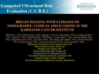
Computed Ultrasound Risk Evaluation (C.U.R.E.). BREAST IMAGING WITH ULTRASOUND TOMOGRAPHY: CLINICAL APPLICATIONS AT THE KARMANOS CANCER INSTITUTE.

E N D
Computed Ultrasound Risk Evaluation (C.U.R.E.) BREAST IMAGING WITH ULTRASOUND TOMOGRAPHY: CLINICAL APPLICATIONS AT THE KARMANOS CANCER INSTITUTE Neb Duric1,2, Ph.D., Peter Littrup1,2, M.D., Cuiping Li1,2, Ph. D., Carri Glide1,2, Ph.D., Lisa Bey-Knight1,2, R.N., Yang Xu1,2,Ph.D., Steve Schmidt1,2, Olsi Rama1,2, Jason Shen1,2, PhD., Anthony Shields, M.D., Ph.D., Ding Wang, M.D., Ph.D., Lou Poulo3, M.Sc., Terry Kling4, Alex Babkin5, Ph.D.,Roman Pevzner5, Ph.D.,Lianjie Huang6, Ph.D., Francesco Simonetti7, Ph.D.,Ivana Jovanovic8 , M.Sc. 1Barbara Ann Karmanos Cancer Institute, Detroit, MI 2Wayne State University, Detroit, MI 3Analogic Corp, Boston, MA 4Sound Technology, State College, PA 5Groupvelocity LLC, Albuquerque, NM 6Los Alamos National Laboratory, Los Alamos, NM 7Imperial College, London, England 8Ecole Polytechnique Federale de Lausanne, Switzerland
C.U.R.E.: Motivation • Mammography has reduced mortality rate by up to 30% Gold standard for screening BUT, problems remain • The false negative problem • ~15% false negative rate missed cancers • Clinical Solution: Improved sensitivity The false positive problem ~80% diagnostic false positive rate high biopsy rate Clinical solution: Improved specificity! • Treatment monitoring problem • Monitoring with imaging difficult / expensive • Clinical solution: Safe, inexpensive whole • breast imaging • Motivation for Computed Ultrasound Risk Evaluation (CURE) Nomass Mammogram: with compression and ionizing radiation.
CURE: The Inverse Problem • Ultrasound Tomography • The Inverse Problem • Measurements of Scattered Ultrasound Field Diagnostic Breast Images • Stability of solution increases with degree of coverage • Quality of reconstruction depends on physics model • Ring geometry to capture total 2-D field • Extraction of multiple tissue parameters for diagnosis • Fast data acquisition to avoid motion artifacts • Low operating frequency • good penetration • closer to 2-D ideal
Water Outlet Patient Bed Water Storage Imaging Tank Electronics Operator’s Display Control Panel Operator’s Desk CURE: Clinical Prototype
CURE: Imaging Tank and Ring Array • A new transducer ring and a new imaging tank have been installed • New cabling added • In plane resolution: 0.5 – 4 mm • Out of plane resolution: 4 mm More than doubled the vertical resolution of the instrument and the signal to noise characteristics.
CURE: Acquisition parameters The numbers • 20cm diameter ring • 256 transmitting and receiving elements • 2.25 MHz frequency • 0.1 seconds to acquire full slice • Transmit with one element receive with all 256 • Repeat the sequence for each transmitting element • 45-60 seconds for a full breast scan
Reflection Sound Speed Attenuation Fusion CURE: Scan Method
CURE Clinical Applications Multiple clinical applications • Diagnostic imaging Adjunct to US and mammography Biopsy reduction Screening • Functional ultrasound imaging • Poor man’s MRI • Assessment of breast cancer risk • Response to therapy • Temperature Monitoring
CURE: Diagnostic Imaging Ultrasound CT – Poor Man’s MRI ?
CURE: Diagnostic Imaging Tomographic 3D MRI-like projections Sound Speed (transmission) images Small, 8-15mm masses (white) are visible in all projections
CURE: Volumetric Imaging 3-D Imaging Operator independent imaging of entire breast volume Exam time < 1 minute CURE imaging: Combines features of mammography, ultrasound and MRI but without compression, ionization or contrast agents. Mammogram: with compression and ionizing radiation.
Multi-modal diagnostics • Cancer identified by its: • high sound speed and attenuation (cyan) • architectural distortion (red). CURE: Diagnostic Imaging R B G + + + ReflectionSound SpeedAttenuation =
CURE: Diagnostic Imaging Clinical Study • Data from ~ 150 patients collected and analyzed • Recruitment at Walt Comprehensive Breast Center at KCI/WSU • CURE exam after mammography, prior to biopsy • CURE results compared against mammography, ultrasound, biopsy and some MRI. • Total recruitment goal: 300
CURE: Diagnostic Imaging Imaging Cancer: Results Detect ~100% cancers. Size range 7 mm – 50 mm. Sound speed sensitivity varies with breast density. Multiple modalities improve sensitivity
CURE: Diagnostic Imaging Imaging Benign Masses: Results Fibroadenoma Hamartoma Cyst Sound Speed Reflection Detect > 90% of benign masses > 5 mm in size Multiple modalities improve differentiation of benign masses from cancer Multiple modalities improve specificity
CURE: Diagnostic Imaging Overall Clinical Implications • Demonstrated ~ 100% sensitivity to masses • Multi-modal imaging visualizes cancers Potential for greater specificity Fast exam times and multi-modal imaging lead to other possible applications
CURE: Breast Density & Cancer Risk • Known correlation between breast density and cancer risk • Currently measured by mammography • Goal: • Replace mammography with CURE • Benefits • Non-ionizing • Non-compressing • Repeatable • Better monitor effect of chemo-preventive agents • Expand access to high risk women Santen, R J, Mansel, R. Benign breast disorders. N Engl J Med, 2005. 353(3): p. 275-85.
CURE: Breast Density & Cancer Risk Ultrasound Percent Density (USPD): As measured with CURE CURE Tomogram Segmented Mammogram Segmented Glide, C., Duric, N., Littrup, P. A Novel Approach to Evaluating Breast Density Utilizing Ultrasound Tomography. Medical Physics. Vol. 34 (2), pp. 744-753. Feb. 2007. • Our study has established equivalence of sound speed and mammography • CURE has potential to replace mammography Potential Application: Chemoprevention and risk screening
CURE: Imaging of Neoadjuvant Patients 18 mm mass 8 mm mass Change in Size and Shape of Mass 08/24/07 10/01/07 Change in Value of Sound Speed of Mass
CURE: Summary • CURE research is exploring multiple areas of clinical applicability • Diagnostic Imaging • Risk Assessment • Assessing treatment response Establish a platform technology • Ongoing Work • Complete clinical study by Aug 2008 • Develop wave based imaging tools for final enhancement of imaging
Acknowledgments • We acknowledge support from following grants: • Sponsor: NCI - Avon, • Improvements in Ultrasound Tomography for Characterization of Breast Masses: Duration: 12/06 – 11//07 • Sponsor: MEDC • Clinical Validation of CURE: A Novel Technology for Improved Breast Cancer Diagnosis: Duration: 02/07 – 08/08 • Komen Foundation grant. • Sound Speed as a Possible Predictor of Breast Cancer Risk.: Duration: 09/07-08/09. • Sponsor: Susan G. Komen foundation • Further reading: Duric, N., Littrup, P., Poulo, L., Babkin, A., Pevzner, R., Holsapple, E., Rama, O., Glide, C. Detection of Breast Cancer With Ultrasound Tomography: First Results with the Computerized Ultrasound Risk Evaluation (C.U.R.E) Prototype. Medical Physics Vol 34 (2), pp. 773-785. Feb 2007.
New scientific publications CURE: Recent Publications • F. Simonetti , L. Huang , N. Duric and O. Rama. Super-resolution ultrasound tomography: A preliminary study with a ring array. Proceedings of SPIE Vol. #6513, 6510-124, 2007. • Pratt, R.G., Huang, L., Duric, N. and Littrup, P.J. Sound-speed and attenuation imaging of breast tissue using waveform tomography of transmission ultrasound data.Proceedings of SPIE Vol. #6513, 6510-174, 2007. • Duric N, Littrup PJ, Rama, O, Holsapple ET. Computerized Ultrasound Risk Evaluation (CURE): First Clinical Results.Acoustical Imaging, Vol 28. MP Andre, Ed., Springer, Dordrecht, 2007, pp. 177-185. • Glide, C., Duric, N., Littrup, P.A Novel Approach to Evaluating Breast Density Utilizing Ultrasound Tomography. Medical Physics. Vol. 34 (2), pp. 744-753. Feb. 2007. • Duric, N., Littrup, P., Poulo, L., Babkin, A., Pevzner, R., Holsapple, E., Rama, O., Glide, C. Detection of Breast Cancer With Ultrasound Tomography: First Results with the Computerized Ultrasound Risk Evaluation (C.U.R.E) Prototype. Medical Physics Vol 34 (2), pp. 773-785. Feb 2007. • Li, C., Huang, L.,, Duric, N., Zhang, H., Rowe, C. An adapted automatic first-arrival picker using the wavelet transform and Akaike information criterion with applications to in-vivo ultrasound breast data. IEEE Transactions on Medical Imaging. Under Review. • F. Simonetti, F., Huang, L., Duric, N. On the spatial sampling of wave fields with circular ring apertures J. Appl. Phys. 101, 083103, 2007 . • Simonetti, F., Huang, L., Duric, N., Rama, O. Imaging beyond the Born approximation: An experimental investigation with an ultrasonic ring array. Physical Review. Accepted..