Download

1 / 6
70 likes | 210 Vues
Dive into the fascinating world of microscopy and uncover the sizes of various specimens from E. coli to red blood cells. Learn how to measure and visualize microscopic objects, including understanding magnification and the field of view when using a microscope. This guide explains how to calculate the size of cells based on field diameter and encourages hands-on exploration of your specimens. Through observation and measurement, see just how tiny these organisms are and what you can discover right under your lens!

E N D
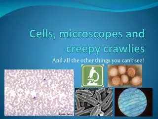
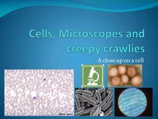
Cells, microscopes and creepy crawlies And all the other things you can’t see!
How small are they really? E. coli: 2-3 μm Influenza virus: 0.130μm OR 130 nm 1000 μm!!! Let’s have a closer look!!! Onion cell: 250-400 μm Red Blood cell: 8 μm
Field of view • When you look in a microscope the circle of light that you see is called the ‘field of view’ • But how do we know how small a cell/specimen really is? • It depends on the magnification • As you increase the magnification the field of view decreases
Measuring a cell! 1,600 μm Field diameter ÷ no. cells ÷ 3 1,600 μm 1000 μm!!! ≈ 533 μm
Measuring a cell! Field diameter ÷ no. cells 4,000 μm 1,600 μm 400 μm Low 4,000 μm ÷ 2 ≈ 2,000 μm ≈ 800 μm 1,600 μm ÷ 2 Medium ≈ 200 μm 400 μm ÷ 2 High
Measuring your own specimens! • We’re going to look at a few specimen • Draw what you see • Write down the magnification • Low = 4 × 10, Medium = 10 × 10, High = 40 × 10 • Guess how many of them you could fit side by side • Use the formula to work out how long / wide your specimen are! • Then we’ll come back to our tables to discuss what we found


![[PDF] DOWNLOAD Creepy Crayon! (Creepy Tales!)](https://cdn7.slideserve.com/12428853/slide1-dt.jpg)


![READ [PDF] Bugs: A Stunning Pop-up Look at Insects, Spiders, and Other Creepy-Crawlies](https://cdn7.slideserve.com/12490851/slide1-dt.jpg)