Outlook
Storet. S 2 (B u ). Q x. Zeaxanthin. Antheraxanthin. S 1 (A g ). Q y. Violaxanthin. S 0 (A g ). Car. BChl. 664 nm. *. *. Chl bulk. Chl-Zea. Zea +*. Chl -*. Chl-Zea. +. SFB Molekulare Bioenergetik -Teilprojekt P27.

Outlook
E N D
Presentation Transcript
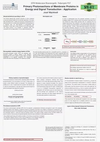
Storet S2 (Bu) Qx Zeaxanthin Antheraxanthin S1 (Ag) Qy Violaxanthin S0 (Ag) Car BChl 664 nm * * Chlbulk Chl-Zea Zea+* Chl-* Chl-Zea + SFB Molekulare Bioenergetik -Teilprojekt P27 Primary Photoreactions of Membrane Proteins in Energy and Signal Transduction: Application Josef Wachtveitl Non-photochemical quenching in LHC-II The recently obtained high resolution structures of LCH-II complexes [1,2] provide detailed insight into the molecular organization of the major lightharvesting protein in plants and should be complemented with a dynamical picture for a fundamental understanding of photosynthesis on a molecular scale. The down-regulation of photosynthetic light harvesting in excess light is mainly achieved via the NPQ-mechanism. The central process is the formation of zeaxanthin (Zea) from violoxanthin (Vio) by deepoxidation in the so called xanthophyll cycle. Xanthophyll cycle Outlook In order to unambiguously prove the proposed mechanism, we want to investigate isolated LHC-II complexes which are enriched in Zea (Zea-LCH-II) and compare the energy transfer dynamics with Vio containing LHC-II by ultrafast spectroscopy. Upon excitation of the lowest excited singlet state of Chl (QY), we plan to record the kinetics in the carotenoid radical cation band in the NIR region (900 - 1100nm). These experiments represent a critical test of the quenching mechanism proposed in [1], which assumes that excitation energy from bulk LHC-II Chl is transferred (very likely via Chl 8) to Zea, where it is trapped and nonradiative dissipation remains as the only decay channel. The qE quenching scheme, which present generation of Zea, upon selective excitation of bulk Chl Qy band at 664 nm is presented below . Energy level diagram of carotenoids and chlorophylls P7 (Kühlbrandt): biochemical experiments and the structural determinations P29 (Dreuw): quantum chemical calculations of NPQ Photosynthetic excitation energy transfer in FCPs: Fucoxanthin-chlorophyll protein (FCP), the peripheral light-harvesting complex in diatoms exhibits a high sequence homology with the LHC-II complex of green plants, but differs substantially in its pigment composition [3], employing carotenoids rather than chlorophylls for the capturing of sunlight. Future plans The ultrafast excitation energy transfer to Chl a followingthe photoexcitation of fucoxanthin (S0S2 transition) shall be studied by vis/near-IR and vis/mid-IR pump-probe experiments. We hope to establish a time scale for the various Car-Chl and Chl-Chl energy transfer reactions in a mode sensitive way. This light harvesting antenna binds Chl a, fucoxanthin and Chl c molecules in a 4:4:1 ratio. Upon excitation of the carotenoid to its S2 state, a significant path of the excitation energy is transferred very fast to Chl a.The energy transfer dynamics that takes place in FCP will be investigated, knowing that the combination of fucoxanthin with a multi-chlorophyll system results into efficient light harvesting. P28 (Büchel): biochemical work and the structural analysis P24 (Hellwig): spectroscopic studies in the far-IR P29 (Dreuw):QM calculation of a model for singlet and triplet energy transfer Primary reactions of proteorhodopsin The structural dynamics of retinal proteins, especially details of the initial photoisomerization of the chromophore is an area of intense efforts. At present, several different models are discussed. The ambiguities are partially due to a lack of experimental time resolution. We therefore plan to study the photodynamics of wild type and mutant proteorhodopsin as well as channelopsin with highest time resolution. As in similar experiments with ultrashort pulses we expect to detect oscillatory contributions, reflecting wave packet dynamics on the excited state potential energy surface. This approach to extract vibrational information from electronic spectra shall be complemented by the recently completed fs mid-IR spectroscopy setup (see P27 report). In 2006 we plan to set up a broadband fs-fluorescence experiment. In collaboration with the Munich group we could already show that this technique also complements fs time resolved absorption experiments and allows detailed observation of the excited state dynamics of retinal proteins. Electron transfer in cytochrome c552: For the photoinitiation of electron transfer in cytochrome c552 we plan the design and the spectroscopic characterization of ruthenium complexes C552/ruthenium. The soluble domain of this protein is ideally suited for these studies, since the structure is known for both redox-states [4]. The ruthenium-labelled cytochrome c552 is also an excellent model system for the study of electron transfer induced folding/unfolding dynamics [5]. This project requires electrochemical techniques and will thus be carried out in close cooperation with: These experiments have already been proposed for the current funding period, but were delayed due to availability problems of the suitable ruthenium reagent and difficulties with the coupling reaction. primary proton donor primary proton acceptor P21 (Mäntele)P24 (Hellwig) P8 (Ludwig). molecular model of proteorhodopsin T. Friedrich et al. (2002) JMB, 321, 821 P1 (Bamberg): electrophysiological studies Does the replacement of Asp97 by Asn confirm the pH dependent measurements (see P27 report)? Photoinduced dynamics in QFR The enzyme quinol:fumarate reductase (QFR) from the anaerobic e-proteobacterium Wolinella succinogenes is part of the anaerobic respiratory system of this organism. It couples the reduction of fumarate to succinate to the oxidation of menaquinol to menaquinone. The three-dimensional structure of the Wollinella succinogenes QFR [6] revealed the exact location of the two heme b groups (bP and bD) within the hydrophobic subunit C. These two hemes are actively involved in the electron transfer reactions driving succinate oxidation and quinone reduction, their proximity and spectral properties make them ideal candidates for optical spectroscopic studies. Layout of the proposed femtosecond fluorescence (Kerr sutter Technique) Using ultrafast spectroscopy with selective excitation of the hemes, we would like to investigate the interactions between hemes bP and bD and the differences in this interaction between the mixed valence and the fully reduced enzyme (DMNH2 vs. dithionite reduced QFR). 1) Standfuss, J., Terwisscha van Scheltinga, A.C., Lamborghini, M. and Kühlbrandt, W. (2005) EMBO J. (in press). 2)Dreuw, A., Fleming, G.R. and Head-Gordon, M. (2003) Phys.Chem. Chem. Phys., 5, 3247–3256. 3) Büchel, C. (2003) Biochemistry, 42, 13027-13034. 4) Harrenga, A., Reincke, B., Rüterjans, H., Ludwig, B. and Michel, H. (2000) J. Mol. Biol., 295, 667-678. 5) Wittung-Stafshede, P., Lee, J.C., Winkler, J.R., Gray, H.B. (1999) Proc. Natl. Acad. Sci. U.S.A., 96, 6587-6590. 6) Lancaster, C.R.D., Kröger, A., Auer, M. and Michel, H. (1999) Nature, 402, 377-385. First time resolved data acquired from a reduced QFR sample by exciting the band of the hems at 387 nm and probing the band transition (530 and 560 nm) P19 (Lancaster): electron transfer studies of the diheme center in QFR