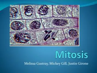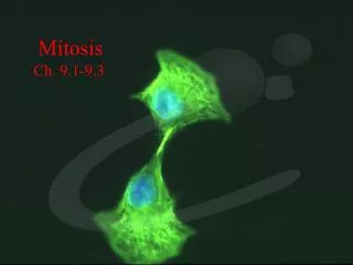
Mitosis
Mitosis. Genes and Proteins. Proteins do the work of the cell: growth, maintenance, response to the environment, reproduction, etc. Proteins are chains of amino acids. The sequence of amino acids in each protein is coded in the DNA as a specific sequence of A, C, G and T bases: a gene.

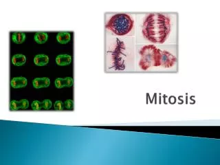
Mitosis
E N D
Presentation Transcript
Genes and Proteins • Proteins do the work of the cell: growth, maintenance, response to the environment, reproduction, etc. • Proteins are chains of amino acids. The sequence of amino acids in each protein is coded in the DNA as a specific sequence of A, C, G and T bases: a gene. • Each gene codes for a different protein. • Key points: • All cells within an organism have the same genes. • What makes cells different from each other is that different genes are turned on and turned off in different cells. • The DNA must be copied and then divided exactly so that each cell gets an identical copy.
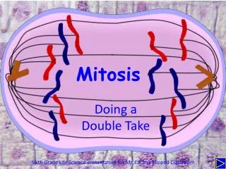
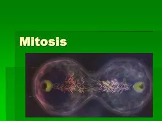
Mitosis • Cells divide to make more cells. While all the other organelles can be randomly separated into the daughter cells, the chromosomes must be precisely divided so that each daughter cell gets exactly the same DNA. • Mitosis is normal cell division, which goes on throughout life in all parts of the body. Meiosis is the special cell division that creates the sperm and eggs, the gametes. We will discuss meiosis separately. • Mitosis and meiosis occur in eukaryotes. Prokaryotes use a different method—”fission” to divide. • Humans have 46 chromosomes, 23 from each parent. Every cell has the same 46 chromosomes Each species has a characteristic number of chromosomes: corn has 20, house flies have 10, chimpanzees have 48.
Chromosomes • The essential part of a chromosome is a single very long strand of DNA. This DNA contains all the genetic information for creating and running the organism. • The DNA is supported and neatly packaged by proteins bound to it. At different times, these proteins cause the DNA to be spread out like spaghetti in a bowl, or tightly condensed into the X-shaped chromosomes we can see in the microscope. • Each chromosome has a central constricted region called a centromere that serves as an attachment point for the machinery of mitosis.
More Chromosomes • Chromosomes exist in 2 different states, before and after they replicate their DNA. Before replication, chromosomes have one chromatid. After replication, chromosomes have 2 sister chromatids, held together at the centromere. Each chromatid is one piece of DNA with its supporting proteins. • In mitosis, the two chromatids of each chromosome separate, with each chromatid going into a daughter cell. • Remember that diploid cells have two copies of each chromosome, one from each parent. These pairs of chromosomes are NOT attached together.
Cell Cycle • Some cells divide constantly: cells in the embryo, skin cells, gut lining cells, etc. Other cells divide rarely or never: only to replace themselves. • Actively dividing cells go through a cycle of events that results in mitosis. Most of the cycle was called “interphase” by the microscopists who first studied cell division. During interphase the cell increases in size, but the chromosomes are invisible. • The 3 stages of interphase are called G1, S, and G2. • The S phase (“Synthesis”) is the time when the DNA is replicated, when the chromosome goes from having one chromatid to having 2 chromatids held together at the centromere. • G1 (“Gap”) is the period between mitosis and S, when each chromosome has 1 chromatid. Cells spend mot of their time in G1: it is the time when the cell grows and performs its normal function. Control of cell division occurs in G1: a cell that isn’t destined to divide stays in G1, while a cell that is to divide enters the S phase. • G2 is the period between S and mitosis. The chromosome have 2 chromatids, and the cell is getting ready to divide.
Machinery of Mitosis • The chromosomes are pulled apart by the spindle, which is made of microtubules. The spindle fibers are attached to each centromere (which is part of the chromosome), and anchored on the other end to a centrosome (which is the organizing center for the spindle). • There are 2 centrosomes, one at each end of the spindle. The chromosomes are lined up between the poles of the spindle. • When the spindle fibers contract, the chromosomes are pulled to the opposing poles. • The cell then divides to separate the two poles. • Stages of mitosis: prophase, metaphase, anaphase, telophase.
Prophase • In prophase, the cell begins the process of division. • 1. The chromosomes condense. The proteins attached to the DNA cause the chromosomes to go from long thin structures to short fat one, which makes them easier to pull apart. • 2. The nuclear envelope disappears. The double membrane that surround the nucleus dissolves into a collection of small vesicles, freeing the chromosomes to use the whole cell for division • 3. The centrosomes move to opposite poles. During interphase, the pair of centrosomes were together just outside the nucleus. In prophase they separate and move to opposite ends of the cell. • 4. The spindle starts to form, growing out of the centrosomes towards the chromosomes.
Metaphase • Metaphase is a short resting period where the chromosomes are lined up on the equator of the cell, with the centrosomes at opposite ends and the spindle fibers attached to the centromeres. Everything is aligned for the rest of the division process to occur.
Anaphase • In anaphase, the centromeres divide. At this point, each individual chromosome goes from: • 1 chromosome with 2 chromatids • to: • 2 chromosomes with one chromatid each. • Then the spindle fibers contract, and the chromosomes are pulled to opposite poles, towards the centrosomes.
Telophase • In telophase the cell actually divides. • The chromosomes are at the poles of the spindle. • The spindle disintegrates • The nuclear envelope re-forms around the two sets of chromosomes. • The cytoplasm is divided into 2 separate cells, the process of cytokinesis.
Cytokinesis • The organelles (other than the chromosomes) get divided up into the 2 daughter cells passively: they go with whichever cell they find themselves in. • Plant and animal cells divide the cytoplasm in different ways. • In plant cells, a new cell wall made of cellulose forms between the 2 new nuclei, about where the chromosomes lined up in metaphase. Cell membranes form along the surfaces of this wall. When the new wall joins with the existing side wall, the 2 cells have become separate. • In animal cells, a ring of actin fibers (microfilaments are composed of actin) forms around the cell equator and contacts, pinching the cell in half.
Summary of Mitosis • Prophase: • Chromosomes condense • Nuclear envelope disappears • centrosomes move to opposite sides of the cell • Spindle forms and attaches to centromeres on the chromosomes • Metaphase • Chromosomes lined up on equator of spindle • centrosomes at opposite ends of cell • Anaphase • Centromeres divide: each 2-chromatid chromosome becomes two 1-chromatid chromosomes • Chromosomes pulled to opposite poles by the spindle • Telophase • Chromosomes de-condense • Nuclear envelope reappears • Cytokinesis: the cytoplasm is divided into 2 cells
Cancer • Cancer is a disease of uncontrolled cell division. It starts with a single cell that loses its control mechanisms due to a genetic mutation. That cell starts dividing without limit, and eventually kills the host. • Normal cells are controlled by several factors. • Normal cells stay in the G1 stage of the cell cycle until they are given a specific signal to enter the S phase, in which the DNA replicates and the cell prepares for division. Cancer cells enter the S phase without waiting for a signal. • Normal cells are mortal. This means that they can divide about 50 times and then they lose the ability to die. This “clock” gets re-set during the formation of the gametes. Cancer cells escape this process of mortality: they are immortal and can divide endlessly. • Normal cells that suffer significant chromosome damage destroy themselves due to the action of a gene called “p53”. Cancer cells either lose the p53 gene or ignore its message and fail to kill themselves.
Cancer Progression • There are many different forms of cancer, affecting different cell types and working in different ways. All start out with mutations in specific genes called “oncogenes”. The normal, unmutated versions of the oncogenes provide the control mechanisms for the cell. The mutations are caused by radiation, certain chemicals (carcinogens), and various random events during DNA replication. • Once a single cell starts growing uncontrollably, it forms a tumor, a small mass of cells. No further progress can occur unless the cancerous mass gets its own blood supply. “Angiogenesis” is the process of developing a system of small arteries and veins to supply the tumor. Most tumors don’t reach this stage. • A tumor with a blood supply will grow into a large mass. Eventually some of the cancer cells will break loose and move through the blood supply to other parts of the body, where they start to multiply. This process is called metastasis. It occurs because the tumor cells lose the proteins on their surface that hold them to other cells.
Cancer Treatment • Two basic treatments: surgery to remove the tumor, and radiation or chemicals to kill actively dividing cells. • It is hard to remove all the tumor cells. Tumors often lack sharp boundaries for easy removal, and metastatic tumors can be very small and anywhere in the body. • Radiation and chemotherapy are aimed at killing actively dividing cells, but killing all dividing cells is lethal: you must make new blood cells, skin cells, etc. So treatment must be carefully balanced to avoid killing the patient. • Chemotherapy also has the problem of natural selection within the tumor. If any of the tumor cells are resistant to the chemical, they will survive and multiply. The cancer seems to have disappeared, but it comes back a few years later in a form that is resistant to chemotherapy. Using multiple drugs can decrease the risk of relapse: it’s hard for a cell to develop resistance to several drugs at the same time.