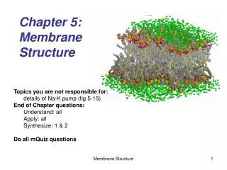
Exploring Cellular Membrane Functions
Learn about the fluid mosaic structure of cell membranes, protein movement within the lipid bilayer, and the impact of different components on membrane fluidity. Understand how membrane proteins function in transport, enzymatic activities, signal transduction, and cell-cell recognition. Explore the synthesis and sidedness of membranes.

Exploring Cellular Membrane Functions
E N D
Presentation Transcript
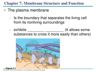
Figure 7.1 Chapter 7: Membrane Structure and Function • The plasma membrane • Is the boundary that separates the living cell from its nonliving surroundings • exhibits ________ _________ (It allows some substances to cross it more easily than others)
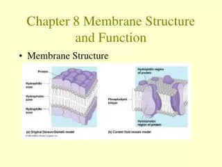
Concept 7.1: Cellular membranes are fluid mosaics of lipids and proteins • ______________ • Are the most abundant lipid in the plasma membrane • Are amphipathic, containing both hydrophobic and hydrophilic regions • The ____ _____ model of membrane structure • States that a membrane is a fluid structure with a “mosaic” of various proteins embedded in it
Lateral movement (~107 times per second) Flip-flop (~ once per month) (a) Movement of phospholipids Figure 7.5 A The Fluidity of Membranes • Phospholipids in the plasma membrane • Can move within the bilayer
Membrane proteins EXPERIMENT Researchers labeled the plasma membrane proteins of a mouse cell and a human cell with two different markers and fused the cells. Using a microscope, they observed the markers on the hybrid cell. RESULTS Mouse cell Mixed proteins after 1 hour Human cell Hybrid cell + CONCLUSION The mixing of the mouse and human membrane proteins indicates that at least some membrane proteins move sideways within the plane of the plasma membrane. Figure 7.6 • Proteins in the plasma membrane • Can drift within the bilayer
Fluid Viscous Unsaturated hydrocarbon tails with kinks Saturated hydro- Carbon tails (b) Membrane fluidity Figure 7.5 B • The type of hydrocarbon tails in phospholipids • Affects the fluidity of the plasma membrane
Cholesterol Figure 7.5 (c) Cholesterol within the animal cell membrane • The steroid cholesterol • Has different effects on membrane fluidity at different temperatures
Interactive Question • Cite some experimental evidence that shows that membrane proteins drift. • How might the plasma membrane of a plant cell change in response to the cold temperatures of winter?
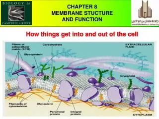
Glycoprotein Carbohydrate Glycolipid EXTRACELLULAR SIDE OF MEMBRANE Microfilaments of cytoskeleton Peripheral protein Cholesterol Integral protein CYTOPLASMIC SIDE OF MEMBRANE Figure 7.7 Membrane Proteins and Their Functions • A membrane • Is a collage of different proteins embedded in the fluid matrix of the lipid bilayer Fibers of extracellular matrix (ECM)
N-terminus C-terminus CYTOPLASMIC SIDE a Helix Figure 7.8 • Integral proteins • Penetrate the hydrophobic core of the lipid bilayer • Are often transmembrane proteins, completely spanning the membrane EXTRACELLULAR SIDE
Peripheral proteins • Are appendages loosely bound to the surface of the membrane
Transport. (left) A protein that spans the membrane may provide a hydrophilic channel across the membrane that is selective for a particular solute. (right) Other transport proteins shuttle a substance from one side to the other by changing shape. Some of these proteins hydrolyze ATP as an energy ssource to actively pump substances across the membrane. (a) ATP (b) Enzymatic activity. A protein built into the membrane may be an enzyme with its active site exposed to substances in the adjacent solution. In some cases, several enzymes in a membrane are organized as a team that carries out sequential steps of a metabolic pathway. Enzymes Signal transduction. A membrane protein may have a binding site with a specific shape that fits the shape of a chemical messenger, such as a hormone. The external messenger (signal) may cause a conformational change in the protein (receptor) that relays the message to the inside of the cell. (c) Signal Receptor Figure 7.9 • An overview of six major functions of membrane proteins
(d) Cell-cell recognition. Some glyco-proteins serve as identification tags that are specifically recognized by other cells. Glyco- protein (e) Intercellular joining. Membrane proteins of adjacent cells may hook together in various kinds of junctions, such as gap junctions or tight junctions (see Figure 6.31). (f) Attachment to the cytoskeleton and extracellular matrix (ECM). Microfilaments or other elements of the cytoskeleton may be bonded to membrane proteins, a function that helps maintain cell shape and stabilizes the location of certain membrane proteins. Proteins that adhere to the ECM can coordinate extracellular and intracellular changes (see Figure 6.29). Figure 7.9
The Role of Membrane Carbohydrates in Cell-Cell Recognition • Cell-cell recognition • Is a cell’s ability to distinguish one type of neighboring cell from another • Membrane carbohydrates • Interact with the surface molecules of other cells, facilitating cell-cell recognition
Synthesis and Sidedness of Membranes • Membranes have distinct inside and outside faces • This affects the movement of proteins synthesized in the endomembrane system
Where are membranes synthesized? • How do the newly made membranes get to the exterior?
1 Transmembrane glycoproteins Secretory protein Glycolipid 2 Golgi apparatus Vesicle 3 Plasma membrane: Cytoplasmic face 4 Extracellular face Transmembrane glycoprotein Secreted protein Membrane glycolipid Figure 7.10 • Membrane proteins and lipids • Are synthesized in the ER and Golgi apparatus ER
The Permeability of the Lipid Bilayer • _________molecules • Are lipid soluble and can pass through the membrane rapidly • ________ molecules • Do not cross the membrane rapidly
Interactive Question • What types of molecules have difficulty crossin the plasma membrane? Why? Ions and larger polar molecules, such as glucose, are impeded by the hydrophobic center of the plasma membrane’s lipid bilayer. Passage through the center of a lipid bilayer is not easy even for small, polar water molecules.
Transport Proteins • Transport proteins • Allow passage of hydrophilic substances across the membrane
(a) Diffusion of one solute. The membrane has pores large enough for molecules of dye to pass through. Random movement of dye molecules will cause some to pass through the pores; this will happen more often on the side with more molecules. The dye diffuses from where it is more concentrated to where it is less concentrated (called diffusing down a concentration gradient). This leads to a dynamic equilibrium: The solute molecules continue to cross the membrane, but at equal rates in both directions. Molecules of dye Membrane (cross section) Equilibrium Net diffusion Net diffusion Figure 7.11 A Concept 7.3: Passive transport is diffusion of a substance across a membrane with no energy investment Diffusion is the tendency for molecules of any substance to spread out evenly into the available space
(b) Diffusion of two solutes. Solutions of two different dyes are separated by a membrane that is permeable to both. Each dye diffuses down its own concen- tration gradient. There will be a net diffusion of the purple dye toward the left, even though the total solute concentration was initially greater on the left side. Equilibrium Net diffusion Net diffusion Net diffusion Equilibrium Net diffusion Figure 7.11 B • Substances diffuse down their concentration gradient, the difference in concentration of a substance from one area to another
Lower concentration of solute (sugar) Higher concentration of sugar Same concentration of sugar Selectively permeable mem- brane: sugar mole- cules cannot pass through pores, but water molecules can Water molecules cluster around sugar molecules More free water molecules (higher concentration) Fewer free water molecules (lower concentration) Osmosis Water moves from an area of higher free water concentration to an area of lower free water concentration Figure 7.12 • Is affected by the concentration gradient of dissolved substances
Water Balance of Cells Without Walls • Tonicity is the ability of a solution to cause a cell to gain or lose water (Has a great impact on cells without walls) • If a solution is _____________ the concentration of solutes is the same as it is inside the cell, and there will be no net movement of water • If a solution is __________, the concentration of solutes is greater than it is inside the cell, and the cell will lose water • If a solution is ____________, the concentration of solutes is less than it is inside the cell, and the cell will gain water
Hypotonic solution Hypertonic solution Isotonic solution (a) Animal cell. An animal cell fares best in an isotonic environ- ment unless it has special adaptations to offset the osmotic uptake or loss of water. H2O H2O H2O H2O Normal Shriveled Lysed Figure 7.13 • Water balance in cells without walls
Hypotonic solution Hypertonic solution Isotonic solution (a) Animal cell. An animal cell fares best in an isotonic environ- ment unless it has special adaptations to offset the osmotic uptake or loss of water. H2O H2O H2O Normal Shriveled Lysed Figure 7.13 • Animals and other organisms without rigid cell walls living in hypertonic or hypotonic environments • Must have special adaptations for osmoregulation H2O
Interactive question • What osmotic problems do freshwater protists face? • What adaptations may help them osmoregulate? • The protists will gain water from their hypotonic environment. • They may have membranes that are less permeable to water and contractile vacuoles that expel excess water.
Water Balance of Cells with Walls • Cell walls help maintain water balance • If a plant cell is turgid • It is in a hypotonic environment • It is very firm (a healthy state in most plants) • If a plant cell is flaccid • It is in an isotonic or hypertonic environment
(b) Plant cell. Plant cells are turgid (firm) and generally healthiest in a hypotonic environ- ment, where the uptake of water is eventually balanced by the elastic wall pushing back on the cell. H2O H2O H2O H2O Turgid (normal) Flaccid Plasmolyzed Figure 7.13 • Water balance in cells with walls
Interactive Question • The ideal osmotic environment for an animal cell is _________ • The ideal osmotic environment for a plant cell is ___________ a. Isotonic b. hypotonic
EXTRACELLULAR FLUID Channel protein Solute CYTOPLASM (a) A channel protein (purple) has a channel through which water molecules or a specific solute can pass. Figure 7.15 Facilitated Diffusion: Passive Transport Aided by Proteins • In facilitated diffusion, transport proteins speed the movement of molecules across the plasma membrane • Channel proteins provide corridors that allow a specific molecule or ion to cross the membrane
Solute Carrier protein (b) A carrier protein alternates between two conformations, moving a solute across the membrane as the shape of the protein changes. The protein can transport the solute in either direction, with the net movement being down the concentration gradient of the solute. Figure 7.15 • Carrier proteins • Undergo a subtle change in shape that translocates the solute-binding site across the membrane
Interactive Question • Why is facilitated diffusion considered passive transport? Although it may speed diffusion, facilitation diffusion is still passive transport because the solute is moving
The Need for Energy in Active Transport • Concept 7.4: Active transport uses energy to move solutes against their gradients • Active transport • Moves substances against their concentration gradient • Requires energy, usually in the form of ATP
2 6 5 1 3 4 [Na+] high [K+] low Na+ Na+ Na+ Na+ Na+ ATP [Na+] low [K+] high P Na+ ADP CYTOPLASM Na+ binding stimulates phosphorylation by ATP. Cytoplasmic Na+ binds to the sodium-potassium pump. Na+ Phosphorylation causes the protein to change its conformation, expelling Na+ to the outside. Na+ Na+ K+ P K+ K+ is released and Na+ sites are receptive again; the cycle repeats. K+ K+ K+ K+ Loss of the phosphate restores the protein’s original conformation. Extracellular K+ binds to the protein, triggering release of the Phosphate group. • The sodium-potassium pump • Is one type of active transport system EXTRACELLULAR FLUID P P i Figure 7.16
Passive transport. Substances diffuse spontaneously down their concentration gradients, crossing a membrane with no expenditure of energy by the cell. The rate of diffusion can be greatly increased by transport proteins in the membrane. Active transport. Some transport proteins act as pumps, moving substances across a membrane against their concentration gradients. Energy for this work is usually supplied by ATP. ATP Diffusion. Hydrophobic molecules and (at a slow rate) very small uncharged polar molecules can diffuse through the lipid bilayer. Facilitated diffusion. Many hydrophilic substances diffuse through membranes with the assistance of transport proteins, either channel or carrier proteins. • Review: Passive and active transport compared Figure 7.17
Maintenance of Membrane Potential by Ion Pumps • Membrane potential • Is the voltage difference across a membrane • An electrochemical gradient • Is caused by the concentration electrical gradient of ions across a membrane
– EXTRACELLULAR FLUID + – ATP + H+ H+ Proton pump H+ + – H+ H+ + – CYTOPLASM + H+ + – • An electrogenic pump • Is a transport protein that generates the voltage across a membrane Figure 7.18
Interactive question • The Na+-K+ pump, the major electrogenic pump in animal cells, exchanges sodium ions for potassium ions, both of which are cations. How does this exchange generate a membrane potential. Three sodium ions are pumped out of the cell for every two potassium ions pumped in, resulting in a net movement of positive charge from the cytoplasm to the extracellular fluid.
– + H+ ATP H+ + – H+ Proton pump H+ – + H+ – + H+ Diffusion of H+ Sucrose-H+ cotransporter H+ – + – Sucrose + Figure 7.19 Cotransport: Coupled Transport by a Membrane Protein • Cotransport is active transport driven by a concentration gradient, and it occurs when active transport of a specific solute indirectly drives the active transport of another solute
Concept 7.5: Bulk transport across the plasma membrane occurs by exocytosis and endocytosis • Large proteins cross the membrane by different mechanisms • In ______________, transport vesicles migrate to the plasma membrane, fuse with it, and release their contents • In ______________, the cell takes in macromolecules by forming new vesicles from the plasma membrane
EXTRACELLULAR FLUID 1 µm CYTOPLASM Pseudopodium Pseudopodium of amoeba “Food” or other particle Bacterium Food vacuole Food vacuole An amoeba engulfing a bacterium via phagocytosis (TEM). In pinocytosis, the cell “gulps” droplets of extracellular fluid into tiny vesicles. It is not the fluid itself that is needed by the cell, but the molecules dissolved in the droplet. Because any and all included solutes are taken into the cell, pinocytosisis nonspecific in the substances it transports. PINOCYTOSIS 0.5 µm Plasma membrane Pinocytosis vesicles forming (arrows) in a cell lining a small blood vessel (TEM). Vesicle • Three types of endocytosis In phagocytosis, a cell engulfs a particle by Wrapping pseudopodia around it and packaging it within a membrane- enclosed sac large enough to be classified as a vacuole. The particle is digested after the vacuole fuses with a lysosome containing hydrolytic enzymes. PHAGOCYTOSIS Figure 7.20
Receptor-mediated endocytosis enables the cell to acquire bulk quantities of specific substances, even though those substances may not be very concentrated in the extracellular fluid. Embedded in the membrane are proteins with specific receptor sites exposed to the extracellular fluid. The receptor proteins are usually already clustered in regions of the membrane called coated pits, which are lined on their cytoplasmic side by a fuzzy layer of coat proteins. Extracellular substances (ligands) bind to these receptors. When binding occurs, the coated pit forms a vesicle containing the ligand molecules. Notice that there are relatively more bound molecules (purple) inside the vesicle, other molecules (green) are also present. After this ingested material is liberated from the vesicle, the receptors are recycled to the plasma membrane by the same vesicle. RECEPTOR-MEDIATED ENDOCYTOSIS Coat protein Receptor Coated vesicle Coated pit Ligand A coated pit and a coated vesicle formed during receptor- mediated endocytosis (TEMs). Coat protein Plasma membrane 0.25 µm
Interactive Question • How is cholesterol, which is used for the synthesis of other steroids and membranes, transported into human cells? • Explain why cholesterol accumulates in the blood of individuals with the disease familial hypercholesteremia. • Human cells are receptor-mediated endocytosis to take in cholesterol. • LDL receptor proteins in the plasma membrane are defective and low-density lipoproteins cannot bind and be transported into the cell.