ATHEROSCLEROSIS Dr. Gehan mohamed
380 likes | 631 Vues
ATHEROSCLEROSIS Dr. Gehan mohamed. Learning objectives. Atherosclerosis Definition of arteriosclerosis and mention the three patterns of it? Mention definition, risk factors, gross morphology, and common sites of atherosclerosis? Discuss microscopic picture of atherosclerosis plaque?

ATHEROSCLEROSIS Dr. Gehan mohamed
E N D
Presentation Transcript
Learning objectives • Atherosclerosis • Definition of arteriosclerosis and mention the three patterns of it? • Mention definition, risk factors, gross morphology, and common sites of atherosclerosis? • Discuss microscopic picture of atherosclerosis plaque? • Mention complications of atherosclerosis? • Discuss pathogenesis of atherosclerotic plaque formation?
ENDOTHELIAL CELLS • single cell-thick, continuous lining of the entire cardiovascular system, collectively called the endothelium. Endothelial structural and functional integrity is fundamental to the maintenance of vessel wall homeostasis and normal circulatory function.
Smooth muscle cells • SMCs are predominant cellular element of the vascular media • SMCs are responsible for vasoconstriction and dilation in response to normal or pharmacologic stimuli. • SMCs are important elements of pathologic processes such as atherosclerosis • Vascular injury/dysfunction stimulates SMCs. They: • migrate from the media to the intima, • Multiply/proliferate as intimal SMCs (In the intima they lose the capacity to contract and gain the capacity to divide). • synthesize collagen, elastin etc and deposit extracellular matrix (ECM).
Arteriosclerosis Arteriosclerosis literally means "hardening of the arteries”. It is a term for thickening and loss of elasticity of arterial walls. Three patterns are recognized: 1)Atherosclerosis, the most frequent and important pattern. 2)Mönckeberg medial calcific sclerosis is characterized by calcific deposits in muscular arteries in older people. 3)Arteriolosclerosis affects small arteries and arterioles, Due to hyaline change of the collagen fibers in the vessel wall causing thickening of it with luminal narrowing and may cause ischemic injury. It Is seen with hypertension and diabetes mellitus.
Atherosclerosis • Atherosclerosis: is characterized by intimal lesions called atheromas, or atheromatous or fibrofatty plaques, which protrude into and obstruct vascular lumens and weaken the underlying media.
The common sites: • abdominal aorta • coronary arteries • the popliteal arteries • the internal carotid arteries • the vessels of the circle of Willis.
Atherosclerosis: Major Risk Factors Potentially modifiable Hyperlipidemia Hypertension Cigarette smoking ,Alcohol Diabetes Obesity Physical inactivity estrogen deficiency Lipoprotein : increase LDL Non-modifiable • Increasing age • Gender : more In males • Family history • Genetic abnormalities
lipoproteins • Some types of lipoproteins promote atheroma formation because they carry lipids to the tissues such as: - Low density lipoproteins (LDLs) - Very low density lipoproteins (VLDLs) -Chylomicrons . • But high density lipoproteins (HDLs) help to protect from atherosclerosis by collecting cholesterol from other lipoproteins and transporting it to liver where it can be metabolized.
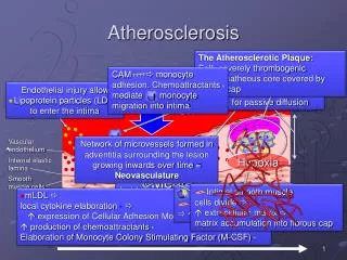
PATHOGENESIS: • response to injury hypothesis it considers atherosclerosis to be a chronic inflammatory response of the arterial wall initiated by injury to the endothelium.
PATHOGENESIS: steps in this thesis are the following: (1)Accumulation of lipoproteins, mainly LDL, with its high cholesterol content, in the vessel wall (2)Chronic endothelial injury caused by smoking ,hypertension. (3)increased permeability of the endothelium to lipoproteins.
PATHOGENESIS: (4)Adhesion of blood monocytes (and other leukocytes) to the endothelium, followed by their migration into the intima and their transformation into macrophages and foamcells (5)Adhesion of platelets (6)Release of factors from activated platelets, macrophages, or vascular cells that cause migration of SMCs from media into the intima
PATHOGENESIS: response to injury hypothesis (7)Proliferation of smooth muscle cells in the intima, and elaboration of extracellular matrix, leading to the accumulation of collagen and proteoglycans (8)Enhanced accumulation of lipidsboth within cells (macrophages and SMCs) and extra cellularly.
How to Make an Atheroma 1.Chronic endothelial “injury” Slide 12.13
How to Make an Atheroma 2 Endothelial dysfunction Monocyte adhesion and emigration
How to Make an Atheroma 3 Macrophage activation Smooth muscle recruitment
5, Well-developed plaque. Slide 12.17
Atherosclerosis: Gross and microscopic morphology • (1)fatty spots are the earliest lesion of atherosclerosis as multiple yellow, flat spots less than 1 mm in diameter .They are composed of lipid-filled foam cells. • (2)Fatty streaksfatty spots coalesce into elongated streaks, 1 cm long or longer. They contain T lymphocytes and extracellular lipid in smaller amounts than in plaques.They are not significantly raised and thus do not cause any disturbance in blood flow.
Fatty streak—a collection of foam cells in the intima Photomicrograph of fatty streak in an experimental hypercholesterolemic rabbit, demonstrating intimal macrophage-derived foam cells ( arrow). Aorta with fatty streaks ( arrows). Slide 12.9
(3) atheroma or atheromatous plaque consists of a raised focal lesion initiating within the intima, having a soft, yellow, core of lipid (mainly cholesterol and cholesterol esters), covered by a firm, white fibrous cap. -size of plaque can reach from 0.3 -1.5 cm so can impinge on the lumen of the artery . -Atherosclerotic lesions usually involve only a partial circumference of the arterial wall ("eccentric" lesions) and are patchy and variable along the vessel length.
Gross views of atherosclerosis in the aorta. A. Mild atherosclerosis composed of fibrous plaques, one of which is denoted by the arrow. B. Severe disease with diffuse and complicated lesions with rupture of the plaque. Slide 12.7
Atherosclerosis: Microscopicaly Atherosclerotic plaques have three principal components: • (1) cells, including smoth muscle cells (SMCs), macrophages, and other leukocytes • (2) Extracelular matrix(ECM), including collagen, elastic fibers, and proteoglycans • (3) intracellular and extracellular lipid . These components occur in varying proportions.
The previously mentioned components are arranged in these layers : • (1) fibrous cap in the top composed of collagen, elastic fibers, and proteoglycans. Beneath and to the side of the cap (the "shoulder") is a cellular area consisting of macrophages, SMCs, and T lymphocytes. • (2) necrotic core Deep to the fibrous cap , containing a disorganized mass of lipid cholesterol clefts, debris from dead cells, foam cells, fibrin, , plasma protein
- Foam cells are large, lipid-laden macrophages derived from blood monocytes, but SMCs can also imbibe lipid to become foam cells. - Typical atheromas contain relatively abundant lipid.
Microscopic picture of atheromatous plaque Major components of well-developed atheromatous plaque: fibrous cap composed of proliferating smooth muscle cells, macrophages, lymphocytes, foam cells, and extracellular matrix. The necrotic core consists of cellular debris, extracellular lipid with cholesterol crystals, and foamy macrophages. Slide 12.6
Histologic features of atheromatous plaque in the coronary artery. A. Overall architecture demonstrating a fibrous cap (F) and a central lipid core (C) with typical cholesterol clefts. The lumen (L) has been moderately narrowed. Note the plaque-free segment of the wall ( arrow). In this section, collagen has been stained blue (Masson trichrome stain). B. Higher-power photograph of a section of the plaque shown in A, stained for elastin ( black) demonstrating that the internal and external elastic membranes are destroyed and the media of the artery is thinned under the most advanced plaque ( arrow). C. Higher-magnification photomicrograph at the junction of the fibrous cap and core showing scattered inflammatory cells, calcification ( broad arrow), and neovascularization ( small arrows). Slide 12.8
Clinical presentation of atherosclerosis • A - asymptomatic • B- symptomatic : a- pain in the area supplied by the partially obstructed blood vessels containing stable atheromatous plaque e.g - coronary arteries angina pectoris - popliteal artery intermittent claudication pain in the leg . - carotid arteries transient ischemic attack of the brain . b- ischemic coagulative necrosis in the area supplied by the completely obstructed blood vessels due to complicated atheromatous plaque.
COMPLICATIONS advanced lesion of atherosclerosis may be complicated by • erosion ,ulceration , rupture, of the luminal surface of atheromatous plaques. (2)Hemorrhageinto a plaque may be initiated by rupture of either the overlying fibrous cap or the thin-walled capillaries that vascularize the plaque. A contained hematoma may expand the plaque or induce plaque rupture
COMPLICATIONS (3)Thrombosisusually occurs on disrupted lesions (those with rupture, ulceration, erosion, or hemorrhage) and may partially or completely occlude the lumen. (4)cholesterol emboli or thromboemboli. may result after rupture of atheromatous plaque that induce thrombus formation which may be fragmented into micro emboli
(5) aneurysm :is abnormal dilatation of the arterial wall Which is induced by atrophy of the underlying media, with loss of elastic tissue, causing weakness, and potential rupture. (6) Calcifications:Atheromas often undergo calcification.
Natural history of atherosclerosis Slide 12.5