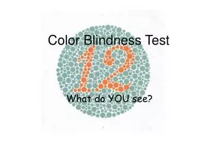
Color Blindness
Color Blindness. By Jessie Wright. Let there be light!!!. Normally, there are three kinds of cones (each one sensitive to a specific range of wavelengths): " red " cones (64%) " green " cones (32%) " blue " cones (2%).


Color Blindness
E N D
Presentation Transcript
Color Blindness By Jessie Wright
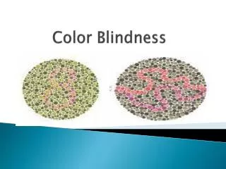
Let there be light!!! Normally, there are three kinds of cones (each one sensitive to a specific range of wavelengths): "red" cones (64%) "green" cones (32%) "blue" cones (2%) The normal human retina contains two kinds of light sensitive cells: the rod cells (active only in low light) and the cone cells (active in normal daylight and responsible for color perception). The different kinds of inherited color blindness result from partial or complete loss of function of one or more of the different cone systems.
Different Types of Color Blindness • Monochromacy: occurs when two or all three of the cone pigments are missing and color and lightness vision is reduced to one dimension. • Total color blindness • Dichromacy: occurs when only one of the cone pigments is missing and color is reduced to two dimensions. • Partial color blindness red-green blue-yellow
Total Color Blindness Also known as rod monochromacy, complete achromatopsia, and typical monochromacy. A rare, non-progressive inability to distinguish any colors as a result of absent or nonfunctioning retinal cones. See everything as white, black, or some shade of gray Typically caused by a missense mutation (a switched amino acid) in the CNGB3 gene.
CYCLIC NUCLEOTIDE-GATED CHANNEL, BETA-3 (CNGB3) Classic achromatopsia results from a complete loss of CNGB3 function. CNGB3 encodes for the beta subunit of the cone cyclic nucleotide-gated cation channel, found photoreceptor plasma membranes. Upon activation by cGMP, it leads to an opening of cation channels which thereby cause a depolarization of rod photoreceptors.This gene is essential for the generation of light-evoked electrical responses in cones. CNGB3 is not required for vital processes outside the visual system.
CNGB3 Cont. The human CNGB3 gene consists of 18 exons distributed over 200 kb of genomic sequence. Gene type: protein coding Domain: similar to ion transport protein Cytogenetic locus: chromosome: 8; Location: 8q21-q22
Amino acid sequence • Length: 809 aa "MFKSLTKVNKVKPIGENNENEQSSRRNEEGSHPSNQSQQTTAQE ENKGEEKSLKTKSTPVTSEEPHTNIQDKLSKKNSSGDLTTNPDPQNAAEPTGTVPEQK EMDPGKEGPNSPQNKPPAAPVINEYADAQLHNLVKRMRQRTALYKKKLVEGDLSSPEA SPQTAKPTAVPPVKESDDKPTEHYYRLLWFKVKKMPLTEYLKRIKLPNSIDSYTDRLY LLWLLLVTLAYNWNCWFIPLRLVFPYQTADNIHYWLIADIICDIIYLYDMLFIQPRLQ FVRGGDIIVDSNELRKHYRTSTKFQLDVASIIPFDICYLFFGFNPMFRANRMLKYTSF FEFNHHLESIMDKAYIYRVIRTTGYLLFILHINACVYYWASNYEGIGTTRWVYDGEGN EYLRCYYWAVRTLITIGGLPEPQTLFEIVFQLLNFFSGVFVFSSLIGQMRDVIGAATA NQNYFRACMDDTIAYMNNYSIPKLVQKRVRTWYEYTWDSQRMLDESDLLKTLPTTVQL ALAIDVNFSIISKVDLFKGCDTQMIYDMLLRLKSVLYLPGDFVCKKGEIGKEMYIIKH GEVQVLGGPDGTKVLVTLKAGSVFGEISLLAAGGGNRRTANVVAHGFANLLTLDKKTL QEILVHYPDSERILMKKARVLLKQKAKTAEATPPRKDLALLFPPKEETPKLFKTLLGG TGKASLARLLKLKREQAAQKKENSEGGEEEGKENEDKQKENEDKQKENEDKGKENEDK DKGREPEEKPLDRPECTASPIAVEEEPHSVRRTVLPRGTSRQSLIISMAPSAEGGEEV LTIEVKEKAKQ"
Mutation… • The genetic basis for achromatopsia is found on chromosome 8 (location 8q21-q22) where there is a recessive point mutation in CNGB3 that changes serine at residue 435 to phenylalanine in a highly conserved site in the S6 membrane-spanning domain.
Bibliography • Bookshelf: • Samir S Deeb, PhD, Arno G Motulsky, MD, “Red-Green Color Vision Defects,” GeneReviews, September 19, 2005. • Berg, Jeremy M. “32.3.5. Rearrangements in the Genes for the Green and Red Pigments Lead to ‘Color Blindness,’” Biochemistry 5th edition, W.H. Freeman and Company, 2002. • OMIM: • “COLORBLINDNESS, PARTIAL, DEUTAN SERIES; CBD” • OMIM ID: 303800 • PubMed: • Harrison, R.; Hoefnagel, D.; Hayward, J. N. “Congenital total color blindness.” Arch. Ophthal. 64: 685-692, 1960. PubMed ID: 13711836 • Botstein, D. “The molecular biology of color vision.”(Editorial) Science 232: 142-143, 1986. PubMed ID: 2937146 • Reyniers, E.; Van Thienen, M.-N.; Meire, F.; De Boulle, K.; Devries, K.; Kestelijn, P.; Willems, P. J. “Gene conversion between red and defective green opsin gene in blue cone monochromacy.”Genomics 29: 323-328, 1995. PubMed ID: 8666378 • Winderickx, J.; Sanocki, E.; Lindsey, D. T.; Teller, D. Y.; Motulsky, A. G.; Deeb, S. S. : • “Defective colour vision associated with a missense mutation in the human green visual pigment gene.” Nature Genet. 1: 251-256, 1992. PubMed ID: 1302020