Download
1 / 25
250 likes | 452 Vues
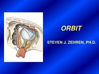
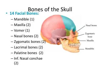
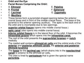
Orbit and the Eye Facial Bones Comprising the Orbit: A. Ethmoid E. Palatine B. Frontal F. Sphenoid C. Lacrimal G. Zygomatic D. Maxillary

E N D
Orbit and the Eye Facial Bones Comprising the Orbit: A. Ethmoid E. Palatine B. Frontal F. Sphenoid C. Lacrimal G. Zygomatic D. Maxillary These bones form a pyramidal-shaped opening below the anterior cranial fossa and in front of the middle cranial fossa. The base of the pyramid is the anterior opening (auditus). The apex is the back end of the orbit and is incomplete due to two openings. superior orbital fissure(opens into middle cranial fossa ) optic canal(also opens into middle cranial fossa) Inferior orbital fissureis in the lateral floor of the orbit. It becomes the infraorbital groovewhich opens into theinfraorbital canal. The roof of the orbit presents thesupraorbital foramenornotchmedially. The medial wall containsethmoid air cellsand the orbital ends of theanteriorandposterior ethmoid canals, theanterior and posterior ethmoid foramina Thefossafor thelacrimal sac(which drains into in thenasolacrimal canal) is anteriorto the ethmoid air cells. The orbit is lined byperiosteumthat is given the special nameperiorbita.
Contents of the Orbit Periorbita (periosteum) Eyeball Ocular muscles and smooth muscles Cranial nerves II, III, IV, V₁,VI Fat Vessels Lacrimal gland and apparatus
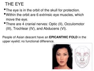
Muscles of the orbit 1. Voluntary Levatorpalpebraesuperioris Superiorrectus Inferiorrectus Medialrectus Lateralrectus Superioroblique Inferioroblique 2. Involuntary Constrictor pupillae Dilator pupillae Superiortarsal
Eyeball (Bulbus Oculi) Consists of three layers (tunics): An outer fibrous tunic, the sclera (white of the eye = 5/6 of eyeball surface) is continuous anteriorly with the transparent cornea. A vascular tunic, the choroid and its anterior extensions forming the ciliarybody and iris with its central opening, the pupil. Smooth muscle in the iris, the constrictor (sphincter) pupillaemuscle, (supplied by parasympathetic fibers from the cilaryganglion, CNIII) reduces the size of the pupil. The pupil is enlarged by the dilatorpupillaemuscle which is supplied by sympatheticfibers. Just behind the sclerocorneal junction, the choroid is thickened and tightly attached to the sclera. The thickened part is the ciliary body. It also contains smooth muscle fibers that form the ciliary muscle. Fibers that suspend the lens, the suspensory ligament of the lens, are attached to the posterior part of the ciliary body. The ciliary muscle (supplied by parasympathetic fibers from the ciliaryganglion, CN III) can contract to release tension on the suspensory ligament of the lens. This allows the elastic lens to assume a more spherical shape and is called accommodation. A tunica interna, the retina, which is the sensory part. The inner surface of the posterior part of the eyeball is called the fundus. The optic nerve leaves the retina at the posterior pole of the eyeball. This area is the opticdisk. The centralarteryoftheretina (a branch of the ophthalmicartery, and an endartery) enters the retina here and branches into fourarterioles (one to each quadrant). One can see the arteries and associated veins with an ophthalmoscope. Enlarged veins or a swollen optic disk (papilladema) can indicate increased intracranial pressure or arterial disease.
The refractive media of the eyeball consist of the cornea, aqueoushumor, lens, and vitreousbody. The aqueoushumor fills the anteriorchamber, which lies between the cornea and the iris and the lens. Glaucoma is a condition of abnormally high pressure in the aqueous humor and can cause blindness. Aqueous humor also fills the posteriorchamber between the iris and the lens. The lens is normally perfectly transparent and is the only refracting medium of the eye whose refracting ability can be varied. A third chamber, the vitreouschamber, lies behind the lens. It is filled with a non-renewing, gelatinous mass, the vitreousbody (humor). A mucous membrane, the conjunctiva, lines the anterior surface of the eyeball up to the periphery of the cornea and the posterior surface of the lids (palpebrae).
Nerves of the orbit Optic (CN II) Oculomotor (CN III) superiordivision inferior division oculomotorrootof ciliaryganglion Trochlear (CN IV) Ophthalmic (CN V₁) frontal lacrimal nasociliary -ganglionic branches to ciliary ganglion -longciliary branches -posteriorethmoidal -anteriorethmoidal -infratrochlear E. Abducens (CN VI) Autonomic Nerves: Parasympathetic 1. Oculomotor (CNIII) – preganglionics to ciliaryganglion; postganglionics to constrictorpupillaemuscle and to ciliarymuscle via shortciliarynerves . 2. Facial (CNVII) – preganglionics to pterygopalatineganglion; postganglionics to lacrimalgland Sympathetic Preganglionics from upper intermediolateralgraycolumn of spinal cord; postganglionics from superiorcervicalganglion to dilatorpupillaemuscle via internalcarotidplexus and nasociliarynerve
Blood Vessels Ophthalmicartery centralarteryoftheretina supraorbital supratrochlear lacrimal Ophthalmicveins 1. Superior and inferior - communicate anteriorly with facialvein and posteriorly with the cavernoussinus Lacrimal Gland and LacrimalApparatus Location of gland: superolateral margin of the orbit. Function: secrete tears to moisten, lubricate, and protect the eyeball Drainage: via punctum and canaliculi at medial corner of eye into the nasolacrimalduct then into inferiormeatus in the nasal cavity Innervation: sensory: lacrimalnerve parasympathetic: preganglionic (greaterpetrosalnerve) branch of the facialnerve synapsing in the pterygopalatineganglion Postganglionic – lacrimal nerve via zygomaticotemporal branch of the maxillary nerve . Eyelids (palpebrae) Tarsal plates are dense fibroelastic tissue covered by skin externally and conjunctiva internally. Orbicularisoculimuscles have a palpebralportion in each lid. Levatorpalpebraesuperiorismuscle inserts into the upper lid. Smooth muscle under this is the superiortarsalmuscle (sympathetic innervation)
Preganglionic parasympathetic fibers of the oculomotor nerve synapse in the ciliary ganglion in the orbit. Sensory fibers from the nasociliary nerve and postganglionic sympathetic fibers from the superior cervical ganglion pass through the ciliary ganglion. All three sets of fibers enter the back of the eyeball as the short ciliary nerves. The parasympathetic fibers innervate the constrictor pupillae muscle and the ciliary muscle (accommodation).