Respiratory Failure/ ARDS
Respiratory Failure/ ARDS. Ian B. Hoffman, MD, FCCP Pulmonary & Critical Care Medicine September 4, 2013.

Respiratory Failure/ ARDS
E N D
Presentation Transcript
Respiratory Failure/ ARDS Ian B. Hoffman, MD, FCCP Pulmonary & Critical Care Medicine September 4, 2013
A 32-year-old man is evaluated for persistent hypoxemia on mechanical ventilation in the intensive care unit. His medical history is significant for paraplegia and a chronic indwelling urinary catheter for neurogenic bladder. He presented to the emergency department 2 days ago with sepsis. At that time, he received piperacillin/tazobactam, normal saline, and vasopressors. He was endotracheallyintubated for decreased level of consciousness. His initial chest radiograph was normal. On physical examination on the second day of hospitalization, temperature is 37.1 °C (98.8 °F), blood pressure is 90/50 mm Hg, pulse rate is 96/min, and respiration rate is 26/min. His need for supplemental oxygen has steadily increased; his oxygen saturation on an FIO2 of 0.8 is 89%. Pulmonary examination reveals bilateral inspiratory crackles. Cardiac examination reveals distant, regular heart sounds. Urine and blood cultures are positive for Escherichia coli. A follow-up chest radiograph shows diffuse bilateral infiltrates without cardiomegaly. Central venous pressure is 8 mm Hg.
Which of the following is the most likely cause of this patient’s hypoxemia? Acute respiratory distress syndrome E. coli pneumonia Heart failure Eosinophilic pneumonia
Respiratory Failure • Any disruption of function of respiratory system – CNS, nerves, muscles, pleura, lungs • Any process resulting in low pO2 or high pCO2 – arbitrarily 50/50 • Acute respiratory failure can be exacerbation of chronic disease or acute process in previously healthy lungs
History • 1940’s – polio, barbiturate OD • 1960’s – blood gas analysis readily available, aware of hypoxemia • 1970’s – decreased hypoxic mortality, increased multiorgan failure (living longer) • 1973 – relationship between resp muscle fatigue and resp failure
Types of Respiratory Failure • Type 1 (nonventilatory) – hypoxemia with or without hypercapnia – disease involves lung itself (i.e, ARDS) • Type 2 – failure of alveolar ventilation – decrease in minute ventilation or increase in dead space (i.e. COPD, drug OD)
Goals of Treatment • Correct hypoxemia or hypercapnia without causing additional complications • Noninvasive ventilation vs. intubation and mechanical ventilation • Goal of mechanical ventilation is NOT necessarily to normalize ABGs
Ventilation–perfusion (V/Q) relationships and associated blood gas abnormalities Shunt
The influence of shunt fraction on the relationship between the inspired oxygen (FiO2) and the arterial PO2 (PaO2).
Ventilatory Failure • Failure of respiratory pump to adequately eliminate CO2 • pCO2 : • VCO2 determined by rate of total body metabolism VCO2 VA
Respiratory Muscles • Acute or acute-on-chronic overloading • COPD, hyperinflation, fatigue • Electrolyte imbalances • Sepsis • Shock • Malnutrition • Drugs • Atrophy related to prolonged mechanical ventilation • Hypothyroidism • Myopathies
What factors leading to respiratory muscle weakness can be reversed? • Reduce respiratory load • treat asthma, COPD, upper airway problems • treat pneumonia, pulm edema, reduce dynamic hyperinflation, drain large pleural effusions, evacuate PTX • Replace K, Mg, PO4, Ca • Treat sepsis • Nutritional support w/o overfeeding • Rest muscles 24-48 hrs, then exercise • Stop aminoglycosides • Rule out hypothyroidism, oversedation, critical illness myopathy/neuropathy
To intubate or not • Decision to mechanically ventilate is clinical • Some criteria: • Decreased level of consciousness (ER always tells us that GCS = 3 and pt tubed to protect airway!) • Vital capacity <15 ml/kg • Severe hypoxemia • Hypercarbia (acute or acute-on-chronic) • Vd/Vt >0.60 • NIF < -25 cm H20
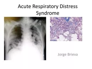
ARDS - Definition • Severe end of the spectrum of acute lung injury • Diffuse alveolar damage • Acute and persistent lung inflammation with increased vascular permeability – inflammatory cytokines • Diffuse infiltrates • Hypoxemia • No clinical evidence of elevated left atrial pressure (PCWP <18 if measured)
ARDS – History/Definitions • 1967 – Ashbaugh described 12 pts with acute respiratory distress, refractory cyanosis, decreased lung compliance, diffuse infiltrates; 7 of the 12 died • 1988 – 4 point lung injury score (level of PEEP, pO2/FiO2, lung compliance, degree of infiltrates) • 1994 – acute onset, bilateral infiltrates, no direct or clinical evidence of LV failure, pO2/FiO2)
Acute onset Bilateral infiltrates c/w pulmonary edema No clinical evidence of left-sided CHF (PCWP <18) paO2/FiO2 ratio <300 Acute onset Bilateral infiltrates c/w pulmonary edema No clinical evidence of left-sided CHF (PCWP <18) paO2/FiO2 ratio <200 1994 American European Consensus Acute Lung Injury ARDS 100/0.40 = 250 100/0.60 = 167
New Definition of ARDS - 2012 • Acute onset (within 7 days of some defined event) • Bilateral infiltrates (on CXR or CT) • No need to exclude heart failure (respiratory failure “not fully explained by CHF”) • Hypoxemia – mild, moderate, severe
ARDS - Incidence • Annual incidence 75 per 100,000 (1977) • 9% of American critical care beds occupied by pts with ARDS
ARDS - Diagnosis • Clinically and radiographically resembles cardiogenic pulmonary edema • PCWP can be misleading – should be normal or low, but can be high • 20% of pts with ARDS may have LV dysfunction
ARDS - Causes • Direct injury to the lung • Indirect injury to the lung in setting of systemic process • Multiple predisposing disorders substantially increase risk • Increased risk with alcohol abuse, chronic lung disease, acidemia
Direct Lung Injury Pneumonia Gastric aspiration Lung contusion Fat emboli Near drowning Inhalation injury Reperfusion injury Indirect Lung Injury Sepsis Multiple trauma Cardiopulm bypass Drug overdose Acute pancreatitis Blood transfusion ARDS - Causes
ARDS - Physiologic Derangements • Inflammatory injury producing diffuse alveolar damage • Alveolar epithelium (eg, aspiration) • Vascular endothelium (eg, sepsis) • Proinflammatory cytokines (TNF, IL-1, IL-8) • Neutrophils recruited – release toxic mediators • Normal barriers to alveolar edema are lost, protein and fluid flow into air spaces, surfactant lost, alveoli collapse; inhomogeneous process • Impaired gas exchange • Decreased compliance • Pulmonary hypertension
ARDS – Features • Severe initial hypoxemia • Increased work of breathing (decreased compliance) – generally a prolonged need for mechanical ventilation • Initial exudative stage • Proliferative stage • resolution of edema, proliferation of type II pneumocytes, squamous metaplasia, collagen deposition • Fibrotic stage
ARDS – Course • Early • Inciting event pulmonary dysfunction (worsening tachypnea, dyspnea, refractory hypoxemia) • Nonspecific labs • CXR – diffuse alveolar infiltrates • Subsequent • Eventual improvement in oxygenation • Continued ventilator dependence • Complications • Large dead space, high minute ventilation requirement • Organization and fibrosis in proliferative phase
ARDS - Complications • Ventilator induced lung injury • Sedation and neuromuscular blockade • Nosocomial infection • Pulmonary emboli • Multiple organ dysfunction
ARDS - Prognosis • Improved survival in recent years – mortality was 50-60% for many years, now 35-40% • Improvements in supportive care, improved mechanical ventilatory management • Early deaths (3 days) usually from underlying cause of ARDS • Later deaths from nosocomial infections, sepsis, MOSF • Respiratory failure only responsible for ~16% of fatalities • Long-term survivors usually show mild abnormalities in pulmonary function (DLCO)
Question 2 • A 63-year-old man with acute respiratory distress syndrome (ARDS) is evaluated in the intensive care unit. He has just been intubated and placed on mechanical ventilation for ARDS secondary to aspiration pneumonia. Before intubation, his oxygen saturation was 78% breathing 100% oxygen with a nonrebreather mask. • On physical examination, temperature is 37.0 °C (98.6 °F), blood pressure is 150/90 mm Hg, and pulse rate is 108/min. His height is 150 cm (59 in) and his weight is 70.0 kg (154.3 lb). Ideal body weight is calculated to be 52.0 kg (114.6 lb). Central venous pressure is 8 cm H2O. Cardiac examination reveals normal heart sounds and no murmurs. Crackles are auscultated in the lower left lung field. The patient is sedated. Neurologic examination is nonfocal. • Mechanical ventilation is on the assist/control mode at a rate of 18/min. Positive end-expiratory pressure is 8 cm H2O, and FIO2 is 1.0.
Which of the following is the most appropriate tidal volume? 300 ml 450 ml 700 ml 840 ml
Ventilatory Goals in ARDS • Provide adequate oxygenation without causing damage related to: • Oxygen toxicity • Hemodynamic compromise • Barotrauma • Alveolar overdistension • Alveolar shear
Mechanical Ventilation in ARDS • Reliable oxygen supplementation • Decrease work of breathing • Increased due to high ventilatory requirements, increased dead space, and decreased compliance • Recruitment of atelectatic lung units • Decreased venous return can help decrease fluid movement into alveolar spaces
Ventilator Induced Lung Injury • Known for decades that high levels of positive pressure ventilation can rupture alveolar units • In 1950’s became known that high FiO2 can produce lung injury • More recently, effects of alveolar overdistension, shearing, cyclical opening and closing have become apparent
Ventilator Induced Lung Injury • Macrobarotrauma • Pneumothorax, interstitial emphysema, pneumomediastinum, SQ emphysema, pneumoperitoneum, air embolism • ? resulting from high airway pressures, or just a marker of severe lung injury • Higher PEEP predicts barotrauma
Ventilator Induced Lung lnjury • Microbarotrauma • Alveolar overinflation exacerbating and perpetuating lung injury – edema, surfactant abnormalities, inflammation, hemorrhage • Less affected lung accommodates most of tidal volume – regional overinflation • Cyclical atelectasis (shear) – adds to injury • Low tidal volume strategy (initial tidal volume 6 ml/kg IBW, plateau pressure <30) – lower mortality
Ventilatory Strategies • Therapeutic target of mechanical ventilation in patients with ARDS has shifted from maintenance of "normal gas exchange” to the protection of the lung from ventilator-induced lung injury • Low tidal volume, plateau pressure <30 peak pressure = large airways plateau pressure = small airways/alveoli • PEEP – enough, not too much • Pressure controlled vs. volume cycled • Prolonging inspiratory time (increase mean airway pressure and improve oxygenation) • APRV • Recent data suggests high frequency oscillation is bad • Permissive hypercapnia • Secondary effect of low tidal volumes • Maintain adequate oxygenation with less risk of barotrauma • Sedation/paralysis often necessary
The only method of mechanical ventilation that has been shown in randomized controlled trials to improve survival in patients with ARDS is low tidal volume ventilation.
ARDS Network Trial NEJM 2000; 342:1301-1308. • Initial tidal volume of 6 ml/kg IBW and plateau pressure <30 vs. Initial tidal volume of 12 ml/kg IBW and plateau pressure <50 • Reduction in mortality of 22% (31% vs 40%)
Ventilator management in patients with acute respiratory distress syndrome or acute lung injury N Engl J Med 2000; 342:1301
Question 3 • A 25-year-old woman is admitted to the intensive care unit (ICU) for a 6-hour history of respiratory distress. She has acute lymphoblastic leukemia and received cytotoxic chemotherapy 2 weeks before ICU admission. She has had fever and leukopenia for 7 days. • On physical examination, she is in marked respiratory distress. Temperature is 39.0 °C (102.2 °F), blood pressure is 110/70 mm Hg, pulse rate is 130/min, and respiration rate is 42/min. Weight is 50.0 kg (110.2 lb). Ideal body weight is calculated as 50.0 kg (110.2 lb). • Acute respiratory distress syndrome is diagnosed. She is intubated and started on mechanical ventilation in the assist/control mode at a rate of 12/min, tidal volume of 300 mL, positive end-expiratory pressure (PEEP) of 5 cm H2O, and FIO2 of 1.0. An arterial blood gas study on these settings shows a pH of 7.47, PCO2 of 30 mm Hg (4.0 kPa), and PO2 of 45 mm Hg (6.0 kPa). Peak airway pressure is 26 cm H2O, and the plateau pressure is 24 cm H2O.
Which of the following is the most appropriate treatment to improve this patient’s oxygenation? Increase PEEP to 10 cm H2O Increase respiratory rate to 18/min Increase tidal volume to 500 ml Start inhaled nitric oxide
PEEP in ARDS • Increases FRC (volume of air remaining in lungs following a normal tidal exhalation) – recruits “recruitable” alveoli, increases surface area for gas exchange • Decreases shunt, improves V/Q matching • No consensus on optimal level of PEEP
ALVEOLI trial NEJM 2004; 351:327-336. • High PEEP vs. low PEEP • Low tidal volume for all (6 ml/kg predicted weight) • Higher PEEP patients had better oxygenation, but no difference in mortality, duration of mechanical ventilation, duration of non-pulmonary organ failure • No benefit from recruitment maneuvers (CPAP 35-40 cm H20 for 30 seconds) – but other studies suggest that recruitment maneuvers do help
Prone Positioning • Thought to improve oxygenation and respiratory mechanics by: • alveolar recruitment • redistribution of ventilation toward dorsal areas resulting in improved V/Q matching • elimination of compression of the lungs by the heart • reduction of parenchymal lung stress and strain
Prone Positioning • Several studies demonstrate improved oxygenation, but no overall reduction in mortality • Greatest benefit of prone positioning occurs in the sickest patients if used early after the diagnosis of ARDS