Download

1 / 4
0 likes | 0 Vues
Discover the hidden link between skin keratinocytes and bone osteoblasts through shared origins, pathways, and disease associations.

E N D
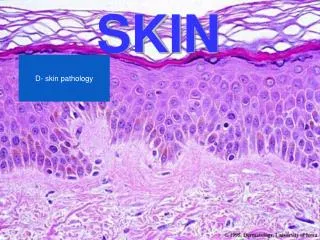
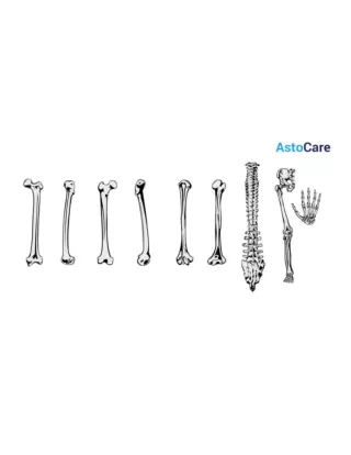
Cross-Talk Between Skin Keratinocytes and Bone Osteoblasts Skin and bones are two different tissue types- soft and hard -serving distinct physiological roles. Their cells, skin keratinocytes and bone osteoblasts, seem disconnected at first glance. But deep exploration has exhibited several surprising similarities between the two cells and their environment. Emerging scientific evidence suggests a connection between the pathologies of both cells, indicating cross-talk between them. The understanding of these mechanisms might provide insights into the pathogenesis and reveal unexpected therapeutic targets. This blog offers a brief overview of the relationship between these cells. Similarities Between Skin Keratinocytes and Bone Osteoblasts Although initially overlooked, scientific investigations have been demonstrating many similarities betweenSkin KeratinocytesandBone Osteoblasts. For instance, they originate from a common precursor- mesenchymal stem cells. Both cells produce collagen, hyaluronic acid, and periostin. Collagen is vital for tissue functioning, defining thickness in the skin and mineral density in bone. Collagen degradation and cross-linking causes thinning in skin and osteoporosis. In fact, the etiologies for collagen breakdown, like age, advanced glycation end-products, and hormonal changes, are identical in both tissues. www.kosheeka.com +91-9654321400
Similarly, calcium is vital for both epithelial barrier and bone homeostasis. Vitamin D deficiency affects both tissues, leading to alopecia and rickets. Studies have suggested that skin-derived biomolecules affect bone functions and bone-derived materials impact the entire body, including skin. The microenvironment of these cells shares similarities as well. Skin epithelium has Langerhans cells to maintain cellular turnover comparable to osteoclasts in bone. Association Between Skin Keratinocytes and Bone Osteoblasts Several skin lesions and bone disorders were found to occur together, reflecting a close relationship between keratinocytes and osteoblasts. Several studies have described the correlation between thin skin and osteoporosis, prompting research into the association between the two cell types. Keratinocytes synthesize vitamin D, which also regulates bone metabolism. A study by Liang et al. showed that keratinocytes secrete cystatin A, which stimulates the proliferation of osteoblasts while inhibiting osteoclast differentiation. With aging, production of cystatin A decreases, which explains the simultaneous occurrence of skin thinning and osteoporosis. Furthermore, Want and BMP signaling pathways in keratinocytes for early skin morphogenesis also participate in bone development. Both cell populations are also linked to stem cell niches and compose a suitable environment for progenitors. Some examples suggesting the correlation between the two are listed below. •Inflammation:Skin disorders such as psoriasis, dermatitis, urticaria, etc., originate due to chronic inflammation. Inflammation also causes bone disorders, hinting at a possible connection between the two cells. Both skin and bone disorders show elevated levels of the same inflammatory mediators, such as TNFα, IL6, IFNϒ, IL7, IL1, IL23, etc. TNFα recruits immune cells in epithelium and promotes osteoclastogenesis in bones, leading to disruption of the epidermal barrier and an increase in bone resorption, respectively. It implies that disorder in either tissue affects the other. Psoriatic arthritis is a key example of this association, where inflammation impacts skin and joints. IL23 cytokine is the central mediator of the disorder. It activates immune cells that, in turn, release IL17, IL22, and TNFα. It is noteworthy that IL17 inhibitors that have therapeutic effects in psoriasis and psoriatic arthritis fail to be effective in rheumatoid arthritis, another inflammatory condition in the joints. •Bisphosphonates:Bisphosphonates are widely employed drugs for bone metastasis and rheumatoid arthritis. However, they raise a serious complication- osteonecrosis of the jaw- a condition marked by loss of soft tissue from jawbone. Research has indicated that the pathways used by bisphosphonates for bone metabolism also impact keratinocytes. For instance, the drug promotes apoptosis in both osteoclasts and keratinocytes. It also increases the expression of BMPs, www.kosheeka.com +91-9654321400
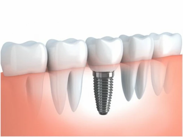
which modulate epithelial cell proliferation and their interaction with fibroblasts, thus affecting skin homeostasis. Bisphosphonates also alter the expression of RANK protein and growth factors, while also suppressing the immune system. Collectively, these pathways impair wound healing by epithelial cells. Concomitant with the role of keratinocytes and osteoblasts, implants are often tested on these cells to evaluate cellular adhesion, viability, and proliferation. •Cholesteatoma:Cholesteatoma is an abnormal skin cyst that develops in the middle ear. In a few cases, it can deteriorate bones and extend to the brain, resulting in meningitis, facial nerve palsy, and abscess. Therefore, researchers have focused on delineating the link between keratinocytes and osteoblasts. In a study by Ishii et al., keratinocytes substantially increase the expression of RANKL in skin fibroblasts. In bone disorders, the RANK-RANKL axis is responsible for activating osteoclasts and causing bone erosion. Therefore, the study implied that fibroblast RANKL induces osteoclast differentiation. It established an indirect interaction of keratinocytes with osteoblasts and indicated that both cells contribute to severe pathologies. •Vitamin D:Vitamin D is a key regulator for both skin and bones. It governs the expression of several genes such as TRPV6, OPG, RANKL, LRP5, etc. Some of these genes modulate signaling pathways in both cells. For example, TRPV6 encodes a calcium ion channel. Its absence decreases bone density and also causes epithelial disorders such as alopecia and dermatitis. Studies indicate that TRPV6 mediates intestinal calcium absorption, vital for bone homeostasis and keratinocyte differentiation. Vitamin D also enhances the Wnt/β-catenin signaling pathway that promotes bone development and induces skin stem cells for hair cycling. Application in Tissue Engineering Tissue engineering aims at replacing damaged organs. In recent years, many different strategies have been employed in this field, among which 3D culture has gained momentum. It employs a scaffold to provide a framework for cultivating cells in a 3D construct. The advances in the technique have divulged that creating a 3D tissue construct that replicates the in vivo function requires more than one cell type. This rationale has been utilized in head and neck construction. They comprise both soft and hard tissues. Thus, researchers have attempted to co-culture keratinocytes and osteoblasts to mimic the in vivo anatomical features. Conclusion Many disorders have indicated a relationship between skin and bone. Although it requires a thorough investigation, early studies definitely suggest a connection between the two. The shared similarities, connecting signaling pathways, co-occurrence of disorders, and www.kosheeka.com +91-9654321400
simultaneous regulation of tissue functions by the same underlying factor hint at the possibility of interaction between them. The exact mechanisms are yet not clear, except for evidence of indirect cross-talk between their cells. These mechanisms can facilitate the development of effective drugs for disorders in both tissues.Kosheekadelivers skin keratinocytes and bone osteoblasts to boost the research on their interaction. Our team also offers customization services with respect to species and donor profile to add precision to your study. FAQ’s Q – What are the functions of keratinocytes? They form the outer layer that acts as a barrier against environmental insults and pathogens. These cells also mount an immune response and repair injury to maintain skin homeostasis. Q – What are the functions of osteoblasts? Osteoblasts are bone architects, that is, they secrete matrix proteins that build bone tissue. They also coordinate with osteoclasts, shaping bone architecture, triggering immune response, and promoting tissue repair. Q – What is the association between keratinocytes and osteoblasts? The simultaneous occurrence of bone and skin disorders suggests an association between the two cells. Studies have shown that they follow similar signaling pathways for physiological functions. Q – Do keratinocytes and osteoblasts interact with each other? The initial studies do not show any direct interaction between these cells. However, an indirect interaction has been established where keratinocytes increase the expression of specific proteins that also regulate osteoblast pathways. www.kosheeka.com +91-9654321400