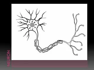
The neuron
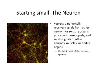
Learn about neurons and how they transmit signals through axon terminals, excitability, ion gradients, resting membrane potential, and action potential stages.


The neuron
E N D
Presentation Transcript
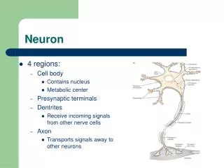
Near its termination each axon divides into fine branches, each of which ends in an axon terminal(ie, synaptic button or botton terminal). The axon terminals contain mitochondria and synaptic vesicles filled with neurotransmitter. These presynaptic structures are the sites where electrical signals are converted into chemical messages for transmission to nearby neurons.
Neurons are able to receive changes in the environment that are transmitted by the nerve pathways to central control organs and than order to the effectors(muscles, glands). Neurons properties are:-excitability-conduction-degeneration and regeneration
Excitability Excitability is the property of a tissue to respond to a stimulus. Stimulus represents any sudden change energy in the environment that alters the permeability of the membrane .
Stimulus must respect the following conditions to produce an excitation: -Intensity; the lowest intensity that produces the response is the threshold intensity. Stimulus with same/above intensity threshold produce excitation, and the stimuli under threshold cannot produce excitation; -Suddenness -Density per unit area -To act a certain period of time. Types of stimuli: electrical, mechanical, chemical, thermal. Excitation is accompanied by changes membrane potential.
The ability of the neuronal membrane to control the movement and concentration of charged particles generates ion gradients with a charge difference across the membrane. The potential difference across the resting membrane is called the resting membrane potential (RMP).
NaCl is found in high concentration outside the neurons, whereas [ K+] is high inside the cell. These ion gradients maintain a constant leakage of NaCl into the cell and a leakage of K+ out. The gradients are maintained by the Na+-K+-pump, which controls the resting membrane potential. Cl - ions distributes passively across most neuronal membranes and contributes little to the resting membrane potential, but they are important for the modulation of incoming signals.
At rest, many K+ -channels are open and K+ moves down its concentration gradient out of the cell, whereby the inside becomes negatively charged(until it is difficult for K+ to leave the cell, and the K+-outflux slows down). The RMP approaches the equilibrium potential for K+.
The following formula, called the Goldman equation, or the Goldman-Hodgkin-Katz equation, gives the calculated membrane potential on the inside of the membrane
Factors participating in the RMP: -Na-K-ATP ase (30% of cell energy is used for its function) -diffusion of ions through the membrane due to unequal permeability for ions, K+ easily crosses the cell membrane but is retained in cells by negative charge given by intracel proteins (negative) whereas membrane permeability for Na+ is lower -Donnan membrane-equilibrium - the presence of intracellular proteins makes the surface negatively charged cell membrane; positive ions (K+) will be retained and anionswill be rejected.
Excitation is a MP change and is expressed electrophysiological by the appearance of action potential (response to action of the stimulus).
The action potential Restingstage. This is the resting membrane potential before the action potential begins. The membrane is said to be “polarized” during this stage because of the –90 mV negative membrane potential that is present.
The action potential Depolarizationstage -the membrane suddenly becomes very permeable to sodium ions, allowing tremendous numbers of positively charged sodium ions to diffuse to the interior of the axon. –by the inflowing positively charged sodium ions the potential rising rapidly in the positive direction. This is called depolarization.
The action potential Repolarizationstage - after the membrane becomes highly permeable to sodium ions, the sodium channels begin to close and the potassium channels open more than normal. -then, rapid diffusion of potassium ions to the exterior re-establishes the normal negative resting membrane potential. This is called repolarization of the membrane.
The action potential is an all-or-none electrical signal, which appears as a positive wave when recording internally. The action potential is conducted with the same shape and size along the whole length of a muscle cell or a nerve fibre.
During the early part of the action potential the cell membrane is completely refractory. A new stimulus, regardless of its size, cannot evoke an action potential. Almost all Na+-channels are inactivated, and will not reopen until the cell membrane is repolarized. This is the absolute refractory period covering most of the peak and lasting until well into the repolarizing phase (ARP).
The action potential During the hyperpolarizing afterpotential, a suprathreshold stimulusis able to trigger a new AP, albeit of smaller amplitude than the first action potential. This period is called the relativerefractory period (RRP). The cell membrane is relatively refractory, because some Na+-channels are voltage-inactivated and at the same time K+-conductance is increased.
The signal conduction is unidirectional, because newly opened Na+ -channels become refractory for a time, when they are inactivated. As these areas are blocked for further depolarization for a time, the depolarization can proceed only in the forward direction towards resting Na+ -channels.
Na+-channels will be recruited in all areas of the membrane, where the threshold potential is exceeded. The Na+-channels behind the peak of the action potential are refractory. This explains why an action potential travels in both directions, when it is evoked in the middle of a nerve.
Special characteristics of signal transmission in (myelinated and unmyelinated) nerve fibers Some fibers are myelinated and the other are unmyelinated. The central core of the fiber is the axon, and the membrane of the axon is the membrane that actually conducts the action potential. The axon is filled in its center with axoplasm Surrounding the axon is a myelin sheath that is often much thicker than the axon itself. About once every 1 to 3 millimeters along the length of the myelin sheath is a nodeof Ranvier.
The myelin sheath consists of 20-300 layers of insulator substance produced by Schwann cells wrapping round the axon. The nodes of Ranvierare the lateral spaces (1 mm wide) between adjacent Schwann cells, which stretch 1-2 mm. Saltatory or leapingconduction occurs, because the action potential is generated only at the nodes.
Even though almost no ions can flow through the thick myelin sheaths of myelinated nerves, they can flow with ease through the nodes of Ranvier. Therefore, action potentials occur only at the nodes; the action potentials are conducted from node to node and this is called saltatory conduction.
That is, electrical current flows through the surrounding extracellular fluid outside the myelin sheath as well as through the axoplasm inside the axon from node to node, exciting successive nodes one after another. Thus, the nerve impulse jumps down the fiber, which is the origin of the term “saltatory.” Saltatory conduction is up to 50 times faster than the conduction through the fastest unmyelinated axons.
Reflex Arc A reflex is an automatic response to a stimulus. Humans use reflex actions in only some of their behaviour, for example controlling the eye's pupil size. Simple reflexes produce rapid involuntary and unconscious responses to a stimulus.
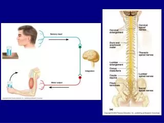
Reflex Arc A reflex arc is the nerve pathway which makes such a fast, automatic response possible. It consists of the following:• A receptor or sense organ,• A sensory neurone (fiber)• A reflex centre (spinal cord or brain)• A motor neurone, and• An effector (muscle or gland).
Sensory receptors are either neurons in the case of vision, smell and cutaneous senses, or modified epithelial cells in the case of auditory, vestibular, smell and taste senses (the special sensory receptors ). Some sensory receptors have characteristics similar to the well-known plasma membrane receptors. Plasma membrane receptors consist of a protein or glycoprotein molecule, an ion channel or a specific enzyme (G-protein).
The adequate stimulus is the stimulus, for which the receptor has a lower energy threshold than for other stimuli (ie, the stimulus to which the receptor is most sensitive). The adequate stimulus for pain receptors is mechanical deformation, extreme temperature or tissue damage.
The stimulation of a receptor elicits a receptor potential (generator potential) that is graded continuously with stimulus intensity. When the stimulus is strong enough to reach the threshold, action potentials (APs) are fired. In neurons the stimulus intensity is coded by the frequency of action potentials. Sensory receptor systems are biological transducers. The depolarisation generates a graded receptor potential forcing a current towards the first node of Ranvier with maintained stimulus.
The receptor potential rapidly decreases (rapid adaptation), because the adequate stimulus is alterations in the deformity rate (vibrations in the range 150-300 Hz). At the first node of Ranvier, a propagating action potential along the axon is released, provided the generator potential is sufficiently large.
Accommodation of sensory receptors (adaptation) is a progressive decrease in firing frequency despite maintained depolarization. The frequency of action potentials from stimulated receptors falls, although the stimulus is maintained at constant strength. Accommodation occurs when a proportion of the voltage-gated Na+-channels is rapidly inactivated by depolarisation, which also opens K+- channels.
Sensory receptors in the nervous system are classified as exteroceptors (located on the body surface), proprioceptors (located in muscles, tendons and joint capsules), interoceptors (located in the viscera), and telereceptors (stimulated by events far from the person).
Sensory receptors are: -mechanoreceptors -thermoreceptors -nociceptors(pain) -electromagnetic( visual) -chemoreceptors
Cutaneous receptors are exteroceptors. Pacinian and Meissner corpuscles are rapidly adapting (dynamic) touch velocity detectors in glabrous skin. In hairy skin, hair-follicle receptors are velocity detectors (they adapt rapidly).
Thermoreceptors are also exteroreceptors and react to temperature changes. -cold-receptors just below the skin surface (200 nm deep). -heat receptors are also located in the skin.
Proprioreceptors • located in muscles, joints and joint capsules • are mechanoreceptors (muscle spindles, Golgi-receptors, Pacinian and Ruffini corpuscles, and free nerve endings) • they provide information for the CNS concerning articular movements, movement velocity and joint position .
Nociceptors or nocireceptors (pain receptors) are responsive to stimuli that potentially cause injury. In the spinal cord nociceptive afferents synapse with secondary neurons in lamina I and II. These sensory neurons ascent in the spinothalamic tracts.
Some signals need to be transmitted to or from the central nervous system extremely rapidly; otherwise, the information would be useless. Nerve fibers come in all sizes between 0.5 and 20 micrometers in diameter—the larger the diameter, the greater the conducting velocity. The range of conducting velocities is between 0.5 and 120 m/sec.
Both conduction velocity and size are used in classification of nerve fibres. The fibres are divided into types A, B and C, based on the three main conduction velocities shown in the record of the compound action potential from a mixed nerve. Type A fibres are the fast conducting myelinated fibres (thick fibres subdivided into alpha, beta , gama , and delta ), type B are preganglionic sympathetic fibres, and type C are the small, unmyelinated fibres.
Synapse In the nervous system, a synapse is a structure that permits a neuron to pass an electrical or chemical signal to another cell (neural or otherwise). There are two major types of synapses: (1) the chemical synapse and (2) the electrical synapse.
Synapse Almost all the synapses used for signal transmission in the central nervous system of the human being are chemical synapses. In these, the first neuron secretes at its nerve ending synapse a chemical substance called aneurotransmitter (or often called simply transmitter substance), and this transmitter in turn acts on receptorproteins in the membrane of the next neuron to excite the neuron, inhibit it, or modify its sensitivity in some other way.
Synapse More than 40 important transmitter substances have been discovered thus far. Some of the best known are acetylcholine, norepinephrine, epinephrine, histamine, gamma-aminobutyric acid (GABA), glycine, serotonin, and glutamate.