Lipids and Proteins
Lipids and Proteins. Lipids A. Fats 1. fatty acids a. saturated b. unsaturated 2. trans fats B. Phosopholipids C. Steroids 1. LDL 2. HDL. Lipids. Lipids are a diverse group of hydrophobic molecules Lipids

Lipids and Proteins
E N D
Presentation Transcript
Lipids A. Fats 1. fatty acids a. saturated b. unsaturated 2. trans fats B. Phosopholipids C. Steroids 1. LDL 2. HDL
Lipids • Lipids are a diverse group of hydrophobic molecules • Lipids • Are the one class of large biological molecules that do not consist of polymers • Share the common trait of being hydrophobic
Fats • Are constructed from two types of smaller molecules, a single glycerol and usually three fatty acids • Vary in the length and number and locations of double bonds they contain
Fats • Vary in the length and number and locations of double bonds they contain
Saturated fatty acids Have the maximum number of hydrogen atoms possible Have no double bonds Stearic acid Figure 5.12 (a) Saturated fat and fatty acid
Oleic acid cis double bond causes bending Figure 5.12 (b) Unsaturated fat and fatty acid • Unsaturated fatty acids • Have one or more double bonds • Mono-unsaturated or polyunsaturated
Monoacylglycerol • Diacyglycerol • Triacylglycerol
In unsaturated fatty acids, there are two ways the pieces of the hydrocarbon tail can be arranged around a C=C double bond. In cis bonds, the two pieces of the carbon chain on either side of the double bond are either both “up” or both “down,” such that both are on the same side of the molecule. In trans bonds, the two pieces of the molecule are on opposite sides of the double bond, that is, one “up” and one “down” across from each other
Naturally-occurring unsaturated vegetable oils have almost all cis bonds, but using oil for frying causes some of the cis bonds to convert to trans bonds. If oil is used only once like when you fry an egg, only a few of the bonds do this so it’s not too bad. However, if oil is constantly reused, like in fast food French fry machines, more and more of the cis bonds are changed to trans until significant numbers of fatty acids with trans bonds build up.
The reason this is of concern is that fatty acids with trans bonds are carcinogenic, or cancer-causing. The levels of trans fatty acids in highly-processed, lipid-containing products such as margarine are quite high, and the government is considering requiring that the amounts of trans fatty acids in such products be listed on the labels.
Phospholipids • Have only two fatty acids • Have a phosphate group instead of a third fatty acid
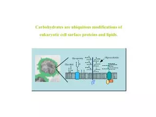
+ CH2 Choline N(CH3)3 CH2 O Phosphate Hydrophilic head – P O O O CH2 CH CH2 Glycerol O O C O C O Fatty acids Hydrophilic head Hydrophobic tails Hydrophobic tails (c) Phospholipid symbol (b) Space-filling model Figure 5.13 (a) Structural formula • Phospholipid structure • Consists of a hydrophilic “head” and hydrophobic “tails”
WATER Hydrophilic head WATER Hydrophobic tail Figure 5.14 • The structure of phospholipids • Results in abilayerarrangement found in cell membranes
H3C CH3 CH3 CH3 CH3 HO Figure 5.15 Steroids • Steroids • Are lipids characterized by a carbon skeleton consisting of four fused rings
One steroid, cholesterol • Is found in cell membranes • Cholesterol is the precursor to our sex hormonesand Vitamin D. Vitamin D is formed by the action of UV light in sunlight on cholesterol molecules that have “risen” to near the surface of the skin. • Our bodies make about 2 g of cholesterol per day, and that makes up about 85% of blood cholesterol, while only about 15% comes from dietary sources.
Lipoproteinsare clusters of proteins and lipids all tangled up together. These act as a means of carrying lipids, including cholesterol, around in our blood. Two main categories of lipoproteins distinguished by how compact/dense they are. 1. LDL or low density lipoproteinis the “bad guy,” being associated with deposition of “cholesterol” on the walls of someone’s arteries. 2. HDL or high density lipoprotein is the “good guy,” being associated with carrying “cholesterol” out of the blood system, and is more dense/more compact than LDL.
FYI only!!! An emulsifying agentis a substance which is soluble in both oil and water, thus enabling the two to mix. A “famous” phospholipid is lecithin which is found in egg yolk and soybeans. Egg yolk is mostly water but has a lot of lipids, especially cholesterol, which are needed by the developing chick. Lecithin is used to emulsify the lipids and hold them in the water as anemulsion. Lecithin is the basis of the classic emulsion known as mayonnaise
Proteins • Proteins have many structures, resulting in a wide range of functions • Proteins do most of the work in cells and act as enzymes • Proteins are made of monomers called amino acids
Table 5.1 • An overview of protein functions
2 2 Substrate binds to enzyme. 1 Active site is available for a molecule of substrate, the reactant on which the enzyme acts. Substrate (sucrose) Enzyme (sucrase) Glucose OH H2O H O Fructose 3 Substrate is converted to products. 4 Products are released. Figure 5.16 • Enzymes • Are a type of protein that acts as a catalyst, speeding up chemical reactions
Amino acids • Are organic molecules possessing both carboxyl and amino groups • Differ in their properties due to differing side chains, called R groups
CH3 CH3 CH3 CH CH2 CH3 CH3 H CH3 H3C CH3 CH2 CH O O O O O H3N+ H3N+ H3N+ H3N+ C H3N+ C C C C C C C C C O– O– O– O– O– H H H H H Valine (Val) Leucine (Leu) Isoleucine (Ile) Glycine (Gly) Alanine (Ala) Nonpolar CH3 CH2 S H2C CH2 O NH CH2 H2N C C CH2 CH2 O– CH2 O O O H H3N+ H3N+ C C C C H3N+ C C O– O– O– H H H Phenylalanine (Phe) Proline (Pro) Methionine (Met) Tryptophan (Trp) Figure 5.17 Twenty Amino Acids • 20 different amino acids make up proteins
OH NH2 O C NH2 O C OH SH CH2 CH3 OH Polar CH2 CH CH2 CH2 CH2 CH2 O O O O O O H3N+ H3N+ H3N+ H3N+ H3N+ H3N+ C C C C C C C C C C C C O– O– O– O– O– O– H H H H H H Glutamine (Gln) Tyrosine (Tyr) Asparagine (Asn) Cysteine (Cys) Serine (Ser) Threonine (Thr) Basic Acidic NH3+ NH2 NH+ O– O –O O CH2 C NH2+ C C NH Electrically charged CH2 CH2 CH2 CH2 CH2 O O H3N+ H3N+ CH2 CH2 C CH2 C C C O O– H3N+ O– CH2 C CH2 C H O H H3N+ O– C C CH2 H O O– H3N+ C C H O– H Lysine (Lys) Histidine (His) Arginine (Arg) Glutamic acid (Glu) Aspartic acid (Asp)
Amino Acid Polymers • Amino acids • Are linked by peptide bonds
Polypeptides • Polypeptides • Are polymers (chains) of amino acids • A protein • Consists of one or more polypeptides
Protein Conformation and Function • A protein’s specific conformation (shape) determines how it functions
Amino acid subunits +H3NAmino end Pro Thr Gly Gly Thr Gly Glu Seu Lys Cys Pro Leu Met Val Lys Val Leu Asp Ala Arg Val Gly Ser Pro Ala Glu Lle Asp Thr Lys Ser Tyr Trp Lys Ala Leu Gly lle Ser Pro Phe His Glu His Ala Glu Val Thr Phe Val Ala Asn lle Thr Asp Ala Tyr Arg Ser Ala Arg Pro Gly Leu Leu Ser Pro Tyr Ser Tyr Ser Thr Thr Ala o Val c Val Glu – Lys o Thr Pro Asn Carboxyl end Figure 5.20 Four Levels of Protein Structure • Primary structure • Is the unique sequence of amino acids in a polypeptide
H H H H H H O O O O O O O H H H H H H R R R R R R R C C C C C C C C C C C C C N N N N N N N N N N N N N C C C C C C C C C C C C C C R R R R R R H H H H H H H O O O O O O O H H H H H H H pleated sheet H O H H Amino acidsubunits C C N N N C C C R H O H H H H H H N N N N N N helix C C O C H H H C C C R R R R R H H C C C C C C O O O O H C R O C C O H C O N N H C C H R H R Figure 5.20 • Secondary structure • Is the folding or coiling of the polypeptide into a repeating configuration • Includes the helix and the pleated sheet
Hydrophobic interactions and van der Waalsinteractions CH CH2 CH2 H3C CH3 OH Polypeptidebackbone H3C CH3 Hydrogenbond CH O HO C CH2 CH2 S S CH2 Disulfide bridge O -O C CH2 CH2 NH3+ Ionic bond • Tertiary structure • Is the overall three-dimensional shape of a polypeptide • Results from interactions between amino acids and R groups
Polypeptidechain Collagen Chains Iron Heme Chains Hemoglobin • Quaternary structure • Is the overall protein structure that results from the aggregation of two or more polypeptide subunits
+H3N Amino end Amino acid subunits helix Review of Protein Structure
Sickle-Cell Disease: A Simple Change in Primary Structure • Sickle-cell disease • Results from a single amino acid substitution in the protein hemoglobin
Normal hemoglobin Sickle-cell hemoglobin Primary structure Primary structure . . . . . . Exposed hydrophobic region Val His Leu Thr Pro Glul Glu Val His Leu Pro Glu Thr Val 5 6 7 3 4 5 6 7 1 2 1 2 3 4 Secondaryand tertiarystructures Secondaryand tertiarystructures subunit subunit Quaternary structure Hemoglobin A Quaternary structure Hemoglobin S Molecules interact with one another tocrystallize into a fiber, capacity to carry oxygen is greatly reduced. Function Molecules donot associatewith oneanother, eachcarries oxygen. Function 10 m 10 m Normal cells arefull of individualhemoglobinmolecules, eachcarrying oxygen Red bloodcell shape Red bloodcell shape Figure 5.21 Fibers of abnormalhemoglobin deform cell into sickle shape.
What Determines Protein Conformation? • Protein conformation Depends on the physical and chemical conditions of the protein’s environment • Temperature, pH, etc. affect protein structure
Denaturation Normal protein Denatured protein Renaturation Figure 5.22 Denaturation is when a protein unravels and loses its native conformation(shape)
The Protein-Folding Problem • Most proteins • Probably go through several intermediate states on their way to a stable conformation • Denaturated proteins no longer work in their unfolded condition • Proteins may be denaturated by extreme changes in pH, temperature, salinity or heavy metals
Correctlyfoldedprotein Polypeptide Cap Hollowcylinder The cap attaches, causing the cylinder to change shape insuch a way that it creates a hydrophilic environment for the folding of the polypeptide. The cap comesoff, and the properlyfolded protein is released. Steps of ChaperoninAction: An unfolded poly- peptide enters the cylinder from one end. Chaperonin(fully assembled) 2 3 1 Figure 5.23 • Chaperonins • Are protein molecules that assist in the proper folding of other proteins
Prions • Prionsare slow-acting, virtually indestructible infectious proteins that cause brain diseases in mammals • Prions propagate by converting normal proteins into the prion version • Scrapie in sheep, mad cow disease, and Creutzfeldt-Jakob disease in humans are all caused by prions
X-raydiffraction pattern Photographic film Diffracted X-rays X-ray beam X-raysource Crystal Nucleic acid Protein (b) 3D computer model (a) X-ray diffraction pattern • X-ray crystallography • Is used to determine a protein’s three-dimensional structure Figure 5.24
Nucleic Acids • Nucleic acids store and transmit hereditary information • Genes • Are the units of inheritance • Program the amino acid sequence of polypeptides • Are made of nucleotide sequences on DNA
The Roles of Nucleic Acids • There are two types of nucleic acids • Deoxyribonucleic acid (DNA) • Ribonucleic acid (RNA)
Deoxyribonucleic Acid • DNA • Stores information for the synthesis of specific proteins • Found in the nucleus of cells
DNA 1 Synthesis of mRNA in the nucleus mRNA NUCLEUS CYTOPLASM mRNA 2 Movement of mRNA into cytoplasm via nuclear pore Ribosome 3 Synthesis of protein Aminoacids Polypeptide Figure 5.25 DNA Functions • Directs RNA synthesis (transcription) • Directs protein synthesis through RNA (translation)
5’ end 5’C O 3’C O O 5’C O 3’C 3’ end OH Figure 5.26 The Structure of Nucleic Acids • Nucleic acids • Exist as polymers called polynucleotides (a) Polynucleotide, or nucleic acid