Lipids and Membranes
Lipids and Membranes. Membranes. All cell membranes share a characteristic trilaminar appearance. Two electron-dense layers separated by a less dense central region. Lipid composition of the plasma membrane and organelle membranes. Stearic and Oleic.

Lipids and Membranes
E N D
Presentation Transcript
Membranes All cell membranes share a characteristic trilaminar appearance. Two electron-dense layers separated by a less dense central region.
Lipid composition of the plasma membrane and organelle membranes
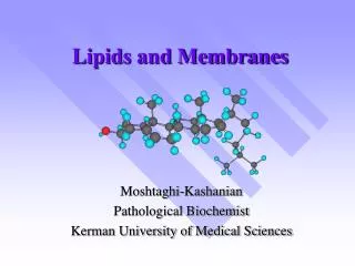
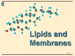
Stearic and Oleic Fatty acids are used as stored forms of energy in cells. A fatty acid is a carboxylic acid with a long unbranched hydrocarbon (aliphatic) chain Lipids are natural products that are insoluble in water. Lipids are involved in energy storage, membrane composition hormone biosynthesis. Lipids are a broad group – fats (triglycerides), sterols, fat-soluble vitamins. Oleic Stearic
Solid Vs Liquid Saturated Vs Unsaturated Simplest fatty acids are unbranched, linear chains of CH2 groups linked by carbon-carbon single bonds with one terminal carboxylic acid group. Saturated indicates that the maximum possible number of hydrogen atoms are bonded to each carbon. An unsaturated fat is a fatty acid in which there is at least one double bond within the fatty acid chain. Monounsaturated contains one double bond, and polyunsaturated contains more than one double bond.
TriGlycerides The simplest lipids constructed from fatty acids are the triacylglycerols. These are also called triglycerides, fats or neutral fats. Triacylglycerols are composed of 3 fatty acids, each in an ester linkage with a single glycerol group. They are nonpolar - and hence water-insoluble - and have lower specific gravity than water. (This is why oil and water don’t mix, and why oil floats on the surface of water) Triglycerides are storage forms of fatty acids. Fatty acids are strong acids and are stored as triacylgycerols Fatty acids are richer in energy than carbohydrates Fatty acids do not interact with water and are stored in anhydrous form (more compact) In human bodies carb can sustain a human for a day while lipids can sustain a human for a couple of weeks
Lipid functions Lipids are used in Fuel storage Hormones Signal transduction messenger Membranes Membrane lipids: Phospholipid Glycolipid Cholesterol
Phospholipid Phospho lipids have a charged group The C1 and C2 of glycerol are esterified with fatty acids C3 OH grp is esterified with phosphoric acid In membranes often the PO4 is further esterified with an alcohol-choline, serine, inositol
Phosphorylcholine Choline is a quaternary saturated amine
GlyceroPhospholipids Cells continually degrade and replace their membrane phospholipids. Enzymes specific for each hydrolyzable bond in a glycerophospholipid exist in lysosomes within cells
Glycolipid Sugar containing lipids Derived from sphingosine Sphingosine has three parts, a three carbon chain with two alcohols an amine and a hydrocarbon chain Sphingomyelin has a sphingosine backbone and a fatty acid is attached to the amine through amide bond. Phosphate is attached through a phosphate ester bond, and through a second phosphate ester bond to choline. Phosphoryl choline is replaced with a sugar
Sterols Sterols are structural lipids present in the membranes of most eukaryotic cells. Cholesterol is the major sterol in animal tissues and is absolutely necessary for membrane biogenesis (especially in children (brain)). All sterols have the 4 ring structure and adopt a cylindrical structure
Vitamin D and Sterol Liver Vitamin D is one of several fat soluble vitamins, compounds that are essential to human health but must be obtained from the diet. Vitamin D forms in the skin by a reaction driven by sunlight; in the liver/kidney, it is converted to a biologically-active hormone that regulates calcium uptake.
Micelle, membrane Vesicle Membrane lipids are amphipathic-hydrophilic polar head and hydrophobic tail
Liposomes Lipid vesicles have low permeability for ions. Na+ or K+ traverse a membrane more slowly than water. Permeability of molecules is correlated with their relative solubility in water or non-polar solvents. Clinical uses of liposomes DNA for gene therapy Drug delivery to tumors (which have more vascularization and therefore more liposomes can be targeted to tumors). Inclusion in liposome membranes of specific cell surface proteins or antibodies against tumor antigens increases specificity of targeting.
Fluid mosaic The fatty acyl chains in the interior of the membrane form a fluid, hydrophobic region. Integral proteins float in this sea of lipid, held by hydrophobic interactions with their non-polar amino acid side chains. Both proteins and lipids are free to move laterally in the plane of the bilayer, but movement of either from one leaflet of the bilayer to the other is restricted. The carbohydrate moieties attached to some proteins and lipids of the plasma membrane are exposed on the extracellular surface of the membrane.
Membrane proteins Integral proteins are embedded in membrane and often span the bilayer Proteins can span the membrane via alpha helices composed on non-polar side chains (bacteriarhodopsin) or beta sheets composed of non-polar side chains (bacterialporin) Peripheral proteins are primarily bound to the head groups by ionic and hydrogen bonds. These polar interactions can be altered by change in pH
Bacteriorhodopsin Bacterialporin
Caveolin forces inward curvature of a membrane. Each caveolin monomer has a central hydrophobic domain and three long-chain acyl groups (red), which hold the molecule to the inside of the plasma membrane. When several caveolin dimers are concentrated in a small region (a raft), they force a curvature in the lipid bilayer
Lipids and proteins are in constant motion via lateral diffusion (raft). Saturated fatty acids favor a rigid membrane Double bonds of unsaturated fatty acids in membranes disrupts ordered packaging of fatty acids and increases mobility of membranes. Cholesterol forms complexes with specific sphingolipids forming thick stable structures called lipid rafts
Transmembrane diffusion- channels In simple diffusion, removal of the hydration shell is highly endergonic, and the energy of activation (ΔG‡) for diffusion through the bilayer is very high A transporter protein reduces the ΔG‡ for transmembrane diffusion of the solute. It does this by forming noncovalent interactions with the dehydrated solute to replace the hydrogen bonding with water and by providing a hydrophilic transmembrane pathway.
Channels animal cell membranes contain a very diverse set of ion channels. Leakage channels are simplest type, permeability is constant. -potassium and chloride channels. Ligand-gated ion channels are channels whose permeability is increased when chemical ligand binds the channel. GABA receptor binds GABA and allows Cl to flow into nerve cells Voltage-gated ion channels are channels whose permeability is influenced by the membrane potential. NMDA receptor allows Ca to flow when electrical potential increases Transmembrane potential is the difference in voltage between the interior and exterior of a cell.
Transport Can be active (energy requiring) or passive
Glucose transport Glucose in blood plasma binds to a stereospecific site on T1 A conformational change from glucoseout • T1 to glucosein • T2, effecting the transmembrane passage of the glucose Glucose is released from T2 into the cytoplasm This is facilitated diffusion
Membrane transport Transporter proteins pump molecules across membranes Small molecules cross a membrane based on a concentration gradient and their solubility in the membrane Lipophilic molecules like cholesterol pass by diffusion. Polar molecules like K+ cannot. Membrane proteins form channels in the membrane that allow K+ to pass through via passive transport. K+ ion entering the channel can pass for 22 A while remaining solvated with water (blue). The pore diameter narrows to 3A (yellow) and K+ shed their water and interact with carbonyl groups of pore amino acids- Gly and Thr Only specific ions can go through specific channels
Primary and secondary active transport In primary active transport the energy released by ATP hydrolysis drives solute movement against an electrochemical gradient In secondary active transport, a gradient of ion- often Na+, has been established by primary active transport. Movement of Na down its electrochemical gradient now provides the energy to drive cotransport of a second solute (S) against its chemical gradient. An electrochemical gradient is a spatial gradient across a membrane of- electrical potential and chemical concentration. Both components are due to ion gradients.
Active Membrane transport Transporter proteins pump molecules across membranes To pump Na+ against a concentration gradient (from areas of low Na+ conc to areas of high Na+ conc) requires energy. Proteins in membrane use energy to move Na+ up a conc gradient via active transport. Active transport of Na+ out of cells and K+ into cells uses a Na+-K+ ATPase. Most ATP in cells is used to perform this transport. This Na+ ion gradient allows muscle cells to be electrically excitable and contract. K+ is a secondary antiporter.
Transport The direction of movement of Na and Ca ions (either inward or outward) depends upon the electrical potential and the chemical gradient for the ions. During ventricular systole (contraction)- the myocytes are depolarized (interior is less negative -60 millivolt) and have a positive membrane potential. The exchanger works such that Na+ leaves the cell and Ca++ enters the cell through the exchanger. Ca++ also enters the cell via Ca channels and Na+ also leaves the cells via the Na/K ATPase pump. Ca++ bind to the myofilaments activating contraction. When the membrane potential is negative (interior is negative -80millivolt), the exchanger reverses action and transports Ca++ out as Na+ enters the cell. Thus during ventricular diasole (resting cells or relaxation of muscle)- the cells are polarized, Ca++ leaves the cell through the exchanger as well as by Ca-ATPase pumps. The flow of Na+ into the muscle and Ca+ out of the muscle, terminates contraction. The exact mechanism by which this exchanger works is unclear. It is known that calcium and sodium can move in either direction via the exchanger. Contraction- more Na outside more Ca inside Relaxation- more Na inside more Ca outside Transmembrane potential is the difference in voltage between the interior and exterior of a cell.
Transport Ca+ Na+-Ca+ Exchanger Ca+ Ca+ Ca+ In heart failure, muscle contraction stops. In heart failure, drugs (digitalis) are given that inhibit the Na+ATPase pump. Inhibition of the pump increases Na+ in cells. This affects the exchanger because it normally relies on low Na+ in the cell to pull more Na+ into the cell simultaneously pumping Ca+ out of the cell. Now since the ATPase is inhibited, Na+ in the cell increases. This reduces the activity of the exchanger. In the presence of the drug, there is a reduced Na+ gradient which results in slower Ca+ expulsion from cell. This leads to increased Ca+ in the cells which causes cells to contract more. CFTR is a Cl- transporter in epithelial cells in the lungs. Reduction of Cl transport leads to CF