
HISTOLOGY 1.10: OSTEOGENESIS
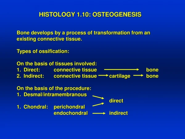
HISTOLOGY 1.10: OSTEOGENESIS. Bone develops by a process of transformation from an existing connective tissue. Types of ossification: On the basis of tissues involved: Direct: connective tissue bone Indirect: connective tissue cartilage bone On the basis of the procedure:

HISTOLOGY 1.10: OSTEOGENESIS
E N D
Presentation Transcript
HISTOLOGY 1.10: OSTEOGENESIS • Bone develops by a process of transformation from an • existing connective tissue. • Types of ossification: • On the basis of tissues involved: • Direct: connective tissue bone • Indirect: connective tissue cartilage bone • On the basis of the procedure: • Desmal/intramembranous • direct • Chondral: perichondral • endochondral indirect
Intramembranous/desmal ossification • The bones of the calvarium develop by this ossification. • The bone forms directly from a layer of mesenchyme in the embryo. • Steps: • Capillaries invade the area • Mesenchymal cells become spherical, form osteoprogenitor cells • Osteoprogenitor cells give rise to osteoblasts, the bone forming cells • Osteoblast synthesize and secrete osteoid, contribute to mineralization • Osteoblast surrounded by the osteoid transform into osteocytes • Formations: • Ossification centers • Bony spicules, trabeculae • Trabecular, spongy or cancellous bone – immature • Replaced by mature lamellar bone
Intramembranous/desmal ossification in light micrographs Low power micrograph showing the „intra- membranous” character 2 1 1 3 2 1: mesenchyme 2: primary bone trabecule 3: osteoclast Arrows show osteoblasts
Endochondral ossification • Bones of the extremities,vertebral column, pelvis, base of skull are formed • by this type of ossification. • The bone is formed initially of hyaline cartilage models that are • replaced by bone. • Steps: • The cartilage model grows more quickly at the end of the long bone • The cartilage cells in the midsection mature and enlarge, release • matrix vesicles, matrix calcifies • Perichondrial mesenchymal cells produce a bony collar around the • diaphysis causing further trouble in the metabolism of the chondrocytes • (direct/perichondral ossification) • A capillary loop accompanied by mesenchymal cells perforates • the bony collar and enters the hypertrophic cartilage • Chondroclasts phagocyte degenerating chondrocytes leaving • pin-shaped calcified remnants of the matrix • Mesenchymal cells differentiate into osteoprogenitor cells which give rise • to osteoblasts laying down the first layers of osteoid onto the surface of • calcified remnants of cartilage matrix
Formation of the bony collar: • A.1. Bony collar formed around the diaphysis • of the degenerating cartilage • A.2. Intact hyaline cartilage at the epiphysis • A.3. Degenerating hyaline cartilage with • hypertrophised chondrocytes and • calcified ground substance • B.1. Ground substance of hyaline cartilage • B.2. Newly divided flattened chondrocytes • B.3. Zone of proliferation • B.4. Zone of degeneration • B.5. Calcium carobonate deposits in • the ground substance
II. Penetration of the capillary loop at the diaphysis A.1. Periosteum around the bony collar A.2. Capillary loop emerging from the periosteum B.1. Osteoblasts on the surface of the bony collar B.2. Osteoclast perforating the bony collar B.3. Bony collar with osteocytes in the lacunae of osteoid B.4. Capillary loop B.5. Multinucleated modified endothel B.6. Mesenchymal cells accompanying the capillary loop
III. Formation of the primary ossification center A.1. Cavity formed by chondroclasts A.2. Blood vessel A.3. Perforation of the bony collar B.1. Bony collar B.2. Hyaline cartilage B.3. Chondrocytes B.4. Periosteum B.5. Capillaries of the periosteum B.6. Perforation site on the bony collar B.7. Blood vessel B.8. Mesenchymal cells B.9. Capillary loop inside the primary bone marrow cavity B.10. Degenerated chondrocyte B.11. Howship’s lacuna B.12. Chondroclast
IV. Zones of the endochondral ossification A.1. Ossification line A.2. Secondary ossification centers B.1. Chondrocyte in mitotic division B.2. Chondroclast phagociting degenerated chondrocytes B.3. Ossification line B.4. Mineralized pin-shaped cartilage ground substance B.5. Osteoblasts on the surface of primary bone trabecule B.6. First layer of osteoid B.7. A primary bone trabecule B.8. Newly embedded osteocyte B.9. A branch of the capillary loop B.I. Zone of resting hyaline cartilage B.II. Zone of proliferation B.III. Zone of maturation B.IV. Zone of degeneration B.V. Zone of ossification 3 2
Endochondral ossification – light microscopy V. IV. III. II. I. Ossification line
Summary of the formation of a long bone with the secondary ossification centers
Growth of the bone: The newly formed bone is transformed into compact or lamellar bone: concurrent periosteal growth and endosteal resorption enlarges the marrow cavity and increases the width of the diaphysis. Epiphyseal plate: disk-shaped intact layer of hyaline cartilage ensure the longitudinal growth of the long bones until puberty. Bone modeling: Primitive woven bone is replaced by lamellar bone. The changes in size and shape during the growth process, it is continuous until adult shape is obtained. Drift: eccentrical growing upon biomechanical demands. Bone remodeling: Does not involve anatomical surfaces, cyclic, and slower procedure. During a lifetime the material of the whole skeleton turn over several times.


