Download
1 / 18
380 likes | 2.89k Vues
Pus (Abscesses, and sinuses) wound, and Burn Cultures. D. M. M. Lab. Pus and wound Culture. Aim of the test To isolate and identify aerobic and anaerobic pathogenic organisms from pus specimen and sensitivity test. Types of specimen

E N D
Pus (Abscesses, and sinuses) wound, and Burn Cultures D. M. M. Lab.
Pus and wound Culture • Aim of the test • To isolate and identify aerobic and anaerobic pathogenic organisms from pus specimen and sensitivity test. • Types of specimen • Swabs from the infected area or aspiration from deep wounds. Swabs in anaerobic transport media for the isolation of anaerobes. • Criteria of specimen rejection • Inappropriate specimen transport device; mislabeled specimen; unlabeled specimen; specimen received after prolonged delay (usually more than 72 hours); specimen received in expired transport media and dried samples.
Pre specimen processing • Who is authorized to order the test • Physician. • Time relapse before processing the sample • According to the type of swab ( recommended within 30 min). • Storage • Maintain specimen swab at room temperature. Do not refrigerate. • Quantity of specimen • Sufficient amount on swab, or aspiration in transport media or syringe ( up to 5 ml of pus ).
Pre specimen processing • Pus and wound swab Specimen collection • No-touching technique: remove bandage with the forceps. • With the forceps take a sponge, dip it in the saline and wash the surface of the wound or ulcers. • Remove the swab from its covering and extend the tip of the swab deep into the wound taking care not to touch the adjacent skin margins. • Remove the stopper from the test tube with transport medium, plunge the swab into the transport medium and replace the stopper.
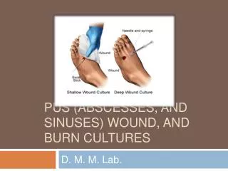
Wound swab Specimen collection The swab should be moved across the wound surface in a zig-zag motion. II. Returning the swab to its container
Pus aspiration Specimen collection • By inserting a needle within a skin lesion, fluid or pus can be aspirated and sent to the laboratory for examination.
Specimen processing for Closed Abscess (wound) NoteAnaerobic cultures should not be routinely set up for superficial-wound specimens or for specimens that have not been submitted in an appropriate anaerobic container.
Specimen processing • Direct smears • gram stain to check the presence or absence and if present the types and predominant organisms. • Culturing • Enrichment and selective media included blood agar, chocolate and MacConkeystreaked and incubated aerobically for 24 hours, and thioglycollatebroth for 24 hours. • In case of suspected anaerobic organisms (closed abscess) another blood agar plate is streaked and incubated anaerobically for 48 hours.
Comments • Cultures from wounds are frequently contaminated with colonized and environmental bacteria, and swab samples often do not reflect the true cause of infection, For this reason the most preferable method of collecting wound specimens is aspirating purulent material from the depths of the wound with a sterile needle and syringe. • The wound margins should be decontaminated as much as possible with swab and alcohol before the material is aspirated. • If Mycobacterium tuberculosis is suspect acid fast stain will be performed.
Burn tissue culture For burn patients, it is important to ascertain the number of organisms present per gram of tissue. Greater than 10^5 CFUs per gram of tissue is considered by some clinicians to be indicative of infection, whereas less than that number may indicate only colonization. Procedure : Cut a piece of tissue measuring several cubic millimeters, aseptically onto a small, pre-weighed, sterile urine cup. Determine the weight of the tissue by subtracting the weight of the aluminum foil from the total weight. Place the specimen and 2 mL of sterile nutrient broth in a sterile tissue grinder; macerate the specimen.
Procedure continue …… 4. Inoculate 0.1 mL of sample to a blood agar plate, in duplicate and anaerobic blood agar plate ( if indicated ), in duplicate. In addition, inoculate 0.01 mL of sample using a calibrated loop to a blood agar plate in duplicate. 5. Spread the inoculum on the plates with sterile glass spreading rod or loop Incubate plates in 5% to 10% carbon dioxide overnight, and count the colonies of bacteria on the plate that contains 30 to 300 CFUs if more than 300 colonies are obtained on broth plated dilution, the factor 300 is used as N for calculations and the result is considered greater than the value. 6. Calculate the number of CFUs per gram of tissue with the following formula: Number of CFUs counted x reciprocal of value of homogenate inoculated (10^-1 or 10^-2) x 2 ( volume of diluent used for tissue homogenization) ÷ weight of tissue in grams.
Post specimen processing • Interfering factors: • Patient on antibiotic therapy. • Improper sample collection. • Result reporting: • Report Gram stain finding as an initial report. • Report the isolated and its sensitivity pattern as a final report. • Turn around time: • Gram stain result should be available half hour after specimen receipt. • Isolation of a possible pathogen can be expected after 2-4 days. • Negative culture will be reported out 1-2 days after the receipt of the specimen.