Download
1 / 84
920 likes | 4.16k Vues
PARANASAL SINUSES- ANATOMY & PATHOLOGY. PRESENTER: DR ASHOK SHARMA JUNIOR RESIDENT II GUIDE: DR P.K. LAMGHARE SIR & DR MOHIT PATIL SIR. PARANASAL SINUSES

E N D
PARANASAL SINUSES- ANATOMY & PATHOLOGY PRESENTER: DR ASHOK SHARMA JUNIOR RESIDENT II GUIDE: DR P.K. LAMGHARE SIR & DR MOHIT PATIL SIR
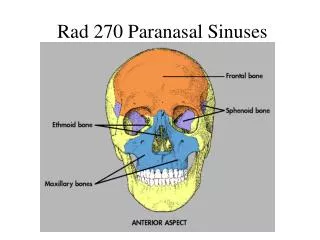
PARANASAL SINUSES • The large, air-filled cavities of the paranasal sinuses are sometimes called the accessory nasal sinuses because they are lined with mucous membrane, which is continuous with the nasal cavity. These sinuses are divided into four groups, according to the bones that contain them: • Maxillary (2) Maxillary (facial) bones • 2. Frontal (usually 2) Frontal (cranial) bones • 3. Ethmoid (many) Ethmoid (cranial) bones • 4. Sphenoid (cranial) bone
Function Histology • They lighten the Facial skeleton • Air-conditioning of the inspired air by providing large surface area over which the air is humidified and warmed. • To provide resonance to voice. • They are lined by Psuedostratified columnar epithelium studded with mucus and serous glands.
Maxillary Sinuses The large maxillary sinuses are paired structures, one of which is located within the body of each maxillary bone. Each maxillary sinus is shaped somewhat like a pyramid on a frontal view. Laterally, they appear more cubic. The average total vertical dimension is between 3 and 4 cm, and the other dimensions are between 2.5 and 3 cm. -drdhiru456@gmail.com
Osteomeatal unit • The osteomeatal unit (OMU) includes the (1) maxillary sinus ostium, (2) ethmoid infundibulum, (3) anterior ethmoid air cells, and (4) frontal recess. • The OMU is the key factor in the pathogenesis of chronic sinusitis.
Frontal Sinuses The frontal sinuses are located between the inner and outer tables of the skull, posterior to the glabella; they rarely become aerated before age 6. The frontal sinuses are always paired and are usually fairly symmetric in size and shape; the frontal sinuses are rarely symmetric.
Ethmoid Sinuses The ethmoid sinuses are contained within the lateral masses or labyrinths of the ethmoid bone. These air cells are grouped into anterior, middle, and posterior collections, but they all intercommunicate. When viewed from the side, the anterior ethmoid sinuses appear to fill the orbits. This occurs because portions of the ethmoid sinuses are contained in the lateral masses of the ethmoid bone, which helps to form the medial wall of each orbit. -drdhiru456@gmail.com
Sphenoid Sinuses The sphenoid sinuses lie in the body of the sphenoid bone directly below the sella turcica. The body of the sphenoid that contains these sinuses is cubic and frequently is divided by a thin septum to form two cavities. This septum may be incomplete or absent entirely, however, resulting in only one cavity.
The Lateral Wall of Nasal Cavity Marked by 3 projections: • Superior concha • Middle concha • Inferior concha • The space below each concha is called a meatus.
The Lateral Wall of Nasal Cavity • Inferior meatus: nasolacrimal duct • Middle meatus: • Maxillary sinus • Frontal sinus • Anterior ethmoid sinuses • Superior meatus: posterior ethmoid sinuses • Sphenoethmoidal recess: sphenoid sinus
LATERAL VIEW Lateral side of the skull lies against the film and x-ray beam is projected perpendicular from the other side. Center CR to a point midway between outer canthus and EAM.
LATERAL POSITION—RIGHT OR LEFT LATERAL: SINUSES Respiration Suspend respiration during exposure.
Structures Shown: • All four paranasal sinus groups are shown.
STRUCTURES SEEN - • ANTERIOR AND POSTERIOR EXTENT OF SPHENOID, FRONTAL AND MAXILLARY SINUSES • SELLA TURCICA • ETHMOID SINUSES • CONDYLE AND NECK OF MANDIBLE
CALDWELL VIEW • A/K/A OCCIPITOFRONTAL VIEW OR NOSE FOREHEAD POSITION
Part Position • Place patient's nose and forehead against upright table with neck extended to elevate the OML 15° from horizontal. A radiolucent support between forehead and upright Bucky or table may be used to maintain this position. CR remains horizontal. (alternate method if Bucky can be tilted 15°.) • Center X-RAY to CR and to nasion, ensuring no rotation. • Align CR horizontal, parallel to floor.
Structures Shown: • Frontal sinuses projected above the frontonasal suture. • Anterior ethmoid air cells visualized lateral to each nasal bone, directly below the frontal sinuses.
STRUCTURES SEEN • FRONTAL SINUSES (SEEN BEST) • ETHMOID SINUSES • MAXILLARY SINUSES • FRONTAL PROCESS OF ZYGOMA AND ZYGOMATIC PROCESS OF FRONTAL BONE • SUPERIOR MARGIN OF ORBIT AND LAMINA PAPYRACEA • SUPERIOR ORBITAL FISSURE
WATER’S VIEW • A.K.A OCCIPITOMENTAL VIEW OR NOSE CHIN POSITION • IT IS TAKEN IN SUCH A WAY THAT NOSE AND CHIN OF THE PATIENT TOUCH THE FILM WHILE X-RAY BEAM IS PROJECTED FROM BEHIND.
Part Position • Extend neck, placing chin and nose against table/film. • Adjust head until MML is perpendicular to film; OML will form a 37° angle with the plane of the film. • Ensure that no rotation or tilt exists. • Center film to CR and to acanthion.
Structures Shown: • Maxillary sinuses with the inferior aspect visualized free from superimposing alveolar processes and petrous ridges, the inferior orbital rim, and an oblique view of the frontal sinuses
STRUCTURES SEEN • Maxillary sinuses (seen best) • Frontal sinuses • Sphenoid sinuses (if the film is taken with open mouth) • Zygoma • Zygomatic arch • Nasal bone • Frontal process of maxilla
SUBMENTOVERTICAL (BASAL) VIEW • THE VIEW IS TAKEN WITH VERTEX NEAR THE FILM AND X-RAY BEAM PROJECTED AT RIGHT ANGLES TO THE FILM FROM THE SUBMENTAL AREA.
Part Position • Raise chin, hyperextend neck if possible until OML is parallel to table/film. • Head rests on vertex of skull. • Ensure no rotation or tilt.
STRUCTURES SEEN • Sphenoid, posterior Ethmoid and Maxillarry sinuses (seen best in that order) • Mandible
Structures Shown: • Sphenoid sinuses, ethmoid sinuses, nasal fossae, and maxillary sinuses.
PARIETOACANTHIAL TRANSORAL PROJECTION: SINUSES Open Mouth Waters Method
Structures Shown: • Maxillary sinuses with the inferior aspect visualized, free from superimposing alveolar processes and petrous ridges, the inferior orbital rim, an oblique view of the frontal sinuses, and the sphenoid sinuses visualized through the open mouth.
Advantages of x-ray imaging in rhinology include:1. Cost effectiveness of the investigation2. Easy availability3. Currently available digital x-ray imaging techniques provide better soft tissue and bone resolution when compared to conventional x-rays.
Disadvantages of conventional radiographs:1. Plain radiographs have a false positive rate of 4% .2. Plain radiographs have false negative rate of more than 30%
BASIC CONCEPTS • CT scans typically obtained for visualizing the paranasal sinus should include coronal and axial (3-mm) cross sections. • Soft tissue and bony windows facilitate evaluation of disease processes and the bony architecture. • The use of intravenous contrast material just prior to scanning can help define soft tissue lesions and delineate vascularized structures, such as vascular tumors. • Contrast-enhanced CT is particularly useful in evaluating neoplastic, chronic, and inflammatory processes.
The CT scan is the GOLD STANDARD investigation in all preoperative cases as it gives detailed bony anatomy of the area and serves as a ‘road map’ for the operating surgeon. • CT scans are best done after a course of antibiotics, so that acute inflammation is not mistaken for chronic mucosal disease.