Download
1 / 51
510 likes | 672 Vues
The Diagnosis of SAH in ED Headache Patients: What Roles for CT Neuroimaging and Lumbar Puncture?. E. Bradshaw Bunney, MD Associate Professor Department of Emergency Medicine University of Illinois at Chicago Our Lady of the Resurrection Medical Center Chicago, IL. Disclosures.

E N D
The Diagnosis of SAH in ED Headache Patients: What Roles for CT Neuroimaging and Lumbar Puncture?
E. Bradshaw Bunney, MDAssociate ProfessorDepartment of Emergency MedicineUniversity of Illinois at ChicagoOur Lady of the Resurrection Medical CenterChicago, IL
Disclosures • AstraZeneca, advisory board • Genentech, speakers bureau • ACEP Scientific Review Committee • Executive Board, Foundation for Education and Research in Neurologic Emergencies
Objectives • Improve screening of patients for SAH • Learn key points in diagnosis, treatment disposition, documentation • Improve outcome of patients with SAH • Further Emergency Medicine practice as it relates to SAH
Patient Clinical History • 47 yo female • Shopping with her husband • Severe, sudden onset of headache • Sat down passed out for 3-5 minutes • Hx of HTN on diuretic
ED Presentation • Vitals: 99.5F, 105, 16, 190/95, 98% RA • Lying still on stretcher with eyes closed • NCAT, Heart, lungs, abdomen normal • “Sore” neck, no clear meningismus • Alert, mild confusion • CN intact, strength 5/5 all 4 ext, sensory intact, DTRs normal, FTN normal
Critical Questions • Who is at risk for SAH? • What symptoms suggest SAH? • How can we best diagnose SAH? • Who requires CT? LP? Angiography? • When should an LP be deferred? • When is “traumatic tap” the likely diagnosis? • When does symptom resolution suggest a benign headache etiology?
SAH Epidemiology • 5% of all strokes • < 1% of all headaches • 50% mortality if not diagnosed • Large risk of litigation
SAH Epidemiology • Majority are traumatic • Non-traumatic • 50% aneurysmal • 15% hypertension • 6% AVM
SAH Presentation • 85% Headache • 40% Nausea and vomiting • Only 15% meningeal signs
SAH Headache • New type of headache • Worst headache of life • Thunderclap – immediate maximal intensity • Warning headache • Sentinel bleed • 15-40% of SAH patients • Typically occur 2 weeks prior to SAH
“Worst Headache of My Life” • N= 107 patients “worst headache” • 20 pts with SAH (19.5%) • 18 of 20 diagnosed by CT (90%) • Two diagnosed: + LP after - CT • NPV of CT = 87/89 = 98% (2% would have SAH)
“Worst Headache” LP Results • Positive LP, Negative CT (n=2) • Tube 1 RBCs: 163,000 median • Tube 4 RBCs: 221,000 median • Negative LP, Negative CT (N = 77) • Tube 1 RBCs: 19 median • Tube 4 RBCs: 0 median
SAH: Risk Stratification • Female • Age > 50 • Exertion • Hypertension • Smoking • Altered consciousness • Neurological deficit • Type of headache
SAH: Diagnostic Tests • CT scan • MRI • Lumbar puncture • Angiography
SAH: CT Scan • Most available • Fast • Most studied • Depend on several factors • Type of scanner • Time since bleeding began • Size of the bleed • Experience of the radiologist
SAH: CT Scan • Sensitivity approaches 100% in 5th generation CT scanners • 3 mm thickness through base of the brain • Within the first 12 hours • 93-95% > 12 hours • Inform the radiologist about possibility of SAH
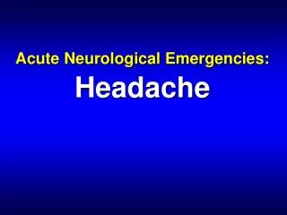
SAH: The Evaluation • How do we evaluate a CT for SAH?
SAH: CT Interpretation • CT evaluation for subarachnoid blood • 1) Inter-hemispheric fissure • 2) Inferior frontal sulci • 3) Third ventricle • 4) Ambient cistern • 5) Sylvian fissure
Inter-hemispheric fissure Sylvian fissure Cistern blood
CT Interpretation: Elevated ICP • CT findings that exclude elevated ICP • Normal cisterns • No obliteration of cistern space • No edema, mass effect, or midline shift • No hydrocephalus
Symptom Resolution • Can headache resolution be used to exclude SAH? • Brings to mind another question…. In a patient who presents to the ED with a headache, can you rule out SAH by clinical evaluation alone?
Symptom Resolution Consider headaches likely benign if: • Low risk SAH patient • No focal neurological findings • Complete symptom resolution with meds that effectively treat migraine and muscle- tension headache (i.e. non-narcotic) • Headache similar to prior headaches
Lumbar Puncture Need Which patients should have a lumbar puncture?
Lumbar Puncture Indications • Moderate to high risk SAH patients following negative CT • Severe, abrupt, thunderclap headache • Focal neurological findings • Unknown CT protocol / interpretive quality • Minimal symptom resolution with meds that effectively treat migraine and muscle- tension headache
Deferred Lumbar Puncture • Is it sometimes reasonable to not perform a lumbar puncture on patients suspected of SAH?
Deferred Lumbar Puncture • Positive CT • Evidence of elevated ICP, edema, mass effect, midline shift, ICH, hydrocephalus • Technically difficult procedure • Critically ill or unstable patient • Coagulopathy
SAH: The Evaluation • How should we interpret CSF results?
Interpreting CSF: RBCs • Likely SAH with: • 10,000-100,000 RBCs or greater • No clearing of RBCs in tube 4 • Consider possible SAH with: • Intermediate RBC count (1,000 – 10,000) • Little RBC clearing by tube 4 • Traumatic tap • 75-90% drop in RBCs from tube 1 to 4
CSF Xanthochromia • Xanthochromia characteristics • Typically > 12 hours from headache onset • Quantitative and qualitative measurements “Read news print test” most often used • Clears after weeks • Oxyhemoglobin = pink, bilirubin = yellow
SAH: The Evaluation • When is angiography indicated?
SAH: Cerebral Angiography • Cerebral angiography indications: • High risk patients with uncertain diagnosis • Interventional radiology available for coiling • Preoperative neurosurgical planning • MRI, MRA, CTA need less well established
SAH: MRI • MRI classically not good at detecting blood • Take longer • Claustrophobia • Not available
SAH: MRI • FLAIR – Fluid-attenuated Inversion Recovery • Detects increase in CSF cellularity and protein • Da Rocha et al. 100% sensitive at detecting SAH up to 15 days after bleed • CT scan 66% sensitive • Small N = 45
Treating SAH Patients • SAH with increased ICP: • Head of the bed at 35 degrees • Mannitol 20% solution 0.25-1.0g per Kg • Hyperventilation to pCO2 30-35 mmHg, temporizing, only if other measures fail • Ventriculostomy • Consider seizure prophylaxis • Nimodopine (vasoconstriction prophylaxis)
ACEP Policy: Acute Headache • Does a response to therapy predict the etiology of an acute headache? • Level C: • Pain response to therapy should not be used as the sole diagnostic criteria in determining the underlying etiology of an acute headache.
ACEP Policy: Acute Headache • In which adults with a headache can an LP be safely performed without neuroimaging? • Level C: Those pts without signs of increased intracranial pressure (ICP) • Papilledema, absent venous pulses • Altered mental status • Focal neurologic deficits
ACEP Policy: Acute Headache • Which patients with an acute headache require neuroimaging? • Level B: • Headache and focal neurologic deficit • Headache of sudden, rapid onset (e.g. SAH) • HIV and new headache • Level C: • > 50 years old, new or different headache
ACEP Policy: Acute Headache • Do patients with “thunderclap” headache need an angiogram after a negative CT and LP? • Level C: • No, outpatient follow-up if: Negative CT, normal opening pressure, and “negative” CSF analysis
ED Patient Management • Pt had a generalized tonic-clonic seizure • Responded to benzodiazepines • Return to normal mental status
ED Diagnostic Evaluation • Non-contrast CT negative • Metabolic, toxicology tests normal • CSF: • Tube 1 = 355,000 RBCs • Tube 4 = 298,000 RBCs • Diagnosis: Subarachnoid Hemorrhage
Patient Outcome • Cerebral angiogram performed • Saccular aneurysm in the posterior communicating artery • Neurosurgical aneurysm clipping • Pt was discharged in one week • No residual neurological deficit
Key Learning Points • SAH needs to be thought of to be diagnosed • Resolution of symptoms does not exclude SAH in all patients • Know the CT technology where you work to be comfortable with the need for LP • When in doubt do the LP