Download
1 / 51
760 likes | 2.58k Vues
Lumbar Disc Herniation and Radiculopathy. KS Hospital Spine Center. Lumbar Spine Motion Segment. Three joint complex Intervertebral disc + 2 facet joint Ligamentous structure, vertebral body. Intervertebral Disc. Hydrostatic, load bearing structure between the vertebral bodies

E N D
Lumbar Disc Herniation and Radiculopathy KS Hospital Spine Center
Lumbar Spine Motion Segment • Three joint complex • Intervertebral disc + 2 facet joint • Ligamentous structure, vertebral body
Intervertebral Disc • Hydrostatic, load bearing structure between the vertebral bodies • Nucleus pulposus + annulus fibrosus • No blood supply • L4-5, largest avascular structure in the body
Nucleus Pulposus • Type II collagen strand + hydrophilic proteoglycan • Water content 70 ~ 90% • Confine fluid within the annulus • Convert load into tensile strain on the annular fibers and vertebral end-plate • Chondrocyte manufacture the matrix component
Annulus Fibrosus • Outer boundary of the disc • More than 60 distinct, concentric layer of overlapping lamellae of type I collagen • Fibers are oriented 30-degree angle to the disc space • Helicoid pattern • Resist tensile, torsional, and radial stress • Attached to the cartilaginous and bony end-plate at the periphery of the vertebra
Vertebral End-Plate • Cartilaginous and osseous component • Nutritional support for the nucleus • Passive diffusion
Facet Joint • Synovial joint • Rich innervation with sensory nerve fiber • Same pathologic process as other large synovial joint • Load share 18% of the lumbar spine
Vital Functions Restricted intervertebral joint motion Contribution to stability Resistence to axial, rotational, and bending load Preservation of anatomic relationship Biochemical Composition Water : 65 ~ 90% wet wt. Collagen : 15 ~ 65% dry wt. Proteoglycan : 10 ~ 60% dry wt. Other matrix protein : 15 ~ 45% dry wt.
Spondylosis • Generalized process of the axial skeleton • Sequence of degenerative change • Start biochemical and cellular level • Manifest biomechanical and morphologic level
Initiating Factor in Degenerative Cascade • Injury to annulus fibrosus • Matrix composition alteration of the nucleus pulposus • Vascularity and permeability change of end-plate • Primary causitive agent?? • The process of disc degeneration is multifactoral
Disc Degeneration Environmental factor Genetic predisposition Normal aging process * Biomechanical stress Degeneration of soft tissue and bone progressive morphologic change
Intervertebral Disc Cellular and Biochemical Change Decrease proteoglycan content Loss of negative charged proteoglycan side chain Water loss within the nucleus pulposus Decrease hydrostatic property Loss of disc height Uneven stress distribution on the annulus
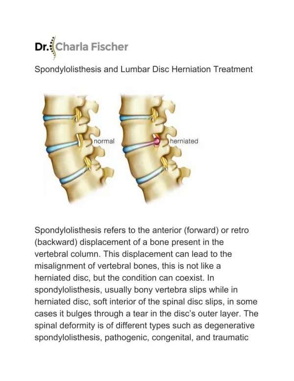
Morphologic Changes bulging of the annulus fibrosus radial tear in-growth of granulation tissue in the annulus annular defect, cleft and fissure cellular necrosis loss of distinction between the nucleus and annulus focal extrusion of disc material Aging Progress disc become more fibrous and disorganized replaced by amorphous fibrocartilage no clear distinction between nucleus and annulus gas formation and vacuum disc sign
Vertebral End-Plate • Become thinner and hyalinized • Decrease permeability • Inhibit nucleus metabolism • Disc space narrowing • Osteophyte formation at the end-plate and annular junction • Marrow change with increased axial loading • Subluxation and instability
Facet Joint Disc height reduction • Facet joint capsule become lax • Increased load transfer to the facet joint • Accelerate degeneration • Joint subluxation, hypertrophy, osteophyte formation ** Primary disc degeneration Secondary change in the posterior facet joint and soft tissue
Lumbar Disc Disease Discogenic Back Pain • Internal Disc Disruption (IDD) • Degenerative Disc Disease (DDD) • Segmental Instability Lumbar Disc Herniation and Radiculopathy
Lumbar Disc Herniation How pain is generated? Inflammatory Biochemical Vascular Mechanical compression
Inflamation • Central role in radiculopathy • Olmarker(1995, spine) Epidural application of autologous nucleus without any pressure Nerve function impairment Axonal injury with significant primary cell damage • Nucleus is totally avascular Perceived as an antigen Intense inflammation response • Application of annulus fibrosus No reduction of nerve conduction velocity
Biochemical Effect • Nuclear herniation Incsease phospolipase A2, prostaglandin E2 cytokine, nitric oxide • Disc herniation and sciatica Neurofilament protein and S-100 increase in CSF Axonal and Schwann's cell damage Mechanical Compression • Local damage and intraneural ischemia
Vascular Pathophysiology * Application of nucleus pulposus to nerve root increase endoneurial pressure decrease blood flow in the dorsal root ganglia compartment syndrome
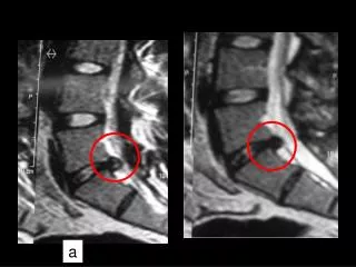
Clinical Anatomy • Disc injury - annular disruption, fissuring, annular defect • Contained herniation Noncontained herniation Extruded Sequestrated • L4-5 and L5-S1 herniation most common - 90% of disc herniation - Great axial load, lordotic shear
History • symptom of disc herniation : acute or gradual • after trauma or without and inciting event • most common 3rd and 4th decade Chief Complain • Pain, radiating from the back or buttock into the leg • Numbness and weakness • Sharp, lancinating, shooting/radiating down the leg posteriorly below the knee • Coughing, Valsalva maneuver increase intracecal pressure increase pain • Sitting position, driving out of lordosis increase intradiscal pressure increase pain
Sciatica - radiating pain down the leg Radiculopathy • radiating pain down the leg as a result of nerve root irritation Back Pain • irritation of the posterior primary ramus - facet capsule, local musculature • sinuvertebral branch - posterior annulus • change in disc loading and shape, biomechanics • loss of viscoelasticity. • 90% of radiating pain have long-standing prior episodic low back pain
Quality of pain and associated symptom • dullache or sharp, stabbing pain? • eletricity, tingling, numbness, shooting down the leg? • any associated weakness? • dose anything make the pain better or worse? • forward flexion or hyperextension exacerbate or relieve pain? • standing more comfortable than sitting? ** Back pain abated when leg pain developed relief of annular tensile stress, nerve root irritation ** Isolated leg pain acute disc extrusion
Differential Diagnosis Vascular claudication • Vascular assessment and flow study • Dorsalis pedis palpation Spinal stenosis • leg pain, dysesthesia, paresthesia, often not dermatomal • pain d/t mechanical compression of spinal canal and foramen • lordosis and axial loading • symptomatic on walking, relief by sitting Thrombophlebitis Metabolic and peripheral neuropathy
Physical Examination Inspection • Old scar, muscle spasm, cutaneous stigma, spinal alignment, loss of lordosis Palpation • Midline, sciatic notch, iliac crest, SI joint, coccyx • Paraspinal tenderness, rigidity • Costovertebral angle, abdomen • Kidney, stone, retroperitoneal abnormality Hip pathology • Patrick test Skin • Temperature and atrophic change
Root Tension Signs • Straight-leg raising : L5, S1 root • Contralateral SLR : sequestrated or extruded disc • Femoral stretching, reverse SLR : L3, L4 root
Diagnostic Test Simple x-ray • Disc space narrowing MRI(magnetic resonance imaging) • Disc pathology, neural structure, musculoligamentous structure • Soft tissue edema, hematoma, intrinsic cord abnormality • Synovial cyst, neurofibroma, perineural cyst • 30% of asymptomatic individual have abnormal MRI CT, Myelography
Nonoperative Treatment • 90% of patient improve with conservative treatment • Short-term rest, NSAID, analgesics, antispamodic medication, exercise • Physical therapy • Oral corticosteroid ** Conservative treatment should continue for 6weeks, before other measure are attempted
Exercise • stretching and strengthening exercise • debate on mechanism of pain relief • protective effect of strong abdominal muscle load share, partially shield the disc from excessive load Physical therapy • heat, cold, massage, ultrasonography • helpful but scientifically not proven
Epidural steroid injection • If leg pain persist beyond 4 weeks • Maximum 3 injection per year • Response vary greatly - Hagen,2002 : short-term effect 40%. no significant long-term effect - Wiesel, 1995 : 82% relief for 1 day, 50% for 2 weeks, 16% for 2mo. - White 1983 : 77% avoid surgery after injection - Carette, 2002 : neither significant functional benefit nor reduction in need for surgery
Indication of Surgery Ideal candidate • history, physical examination, radiographic finding, are consistent with one another • when discrepancy exist, the clinical picture should serve as the principal guide. Absolute surgical indication • cauda equina syndrome • acute urinary retension/incontinence, saddle anesthesia, back/buttock/leg pain, weakness, difficulty walking Relative indication • progressive weakness • no response to conservative treatment