Download
1 / 23
290 likes | 398 Vues
Learn about lumbar disc prolapse, including anatomy of the intervertebral disc, causes, pathophysiology, stages of prolapse, clinical features, cauda equina syndrome, physical examination, and relevant neurological findings.

E N D
Lumbar disc prolapse Done by : Areej Al-Hadidi
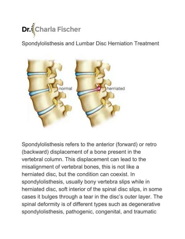
IVD is composed of two components: 1. anulusfibrosus : it is the outer fibrous layer (fibrocartilage ) **It is comressible &tough 2. nucleus pulposus :It is the inner gelatinous layer ** it isn’t compressible **function of IVD: flexibility & shock absorption
Defenetion: Lumbar disc prolapse : herniation of a part of IVD Causes: *It is ay be precipitated by injury or excessive strain *Smoking *Multiparity *Occupation *Obesity *Improper postural habits *Spontaneous age degeneration ***in the majority of cases the back pain is associated with some abnormality of theinter-vertebral discs at the lowest two levels of the spine (L4/5 and (L5/S1
Pathophysiology **When the spine is straight, such as in standing or lying down, internal pressure is equalized on all parts of the discs **when the anterior side of the disc is compressed while sitting or bending forward, the nucleus pulposusget pressed against the tightly stretched and thinned membrane annulus fibrosuson the posterior side (back side) of the disc. the combination of membrane-thinning from stretching and increased ** herniation))internal pressure results in the rupture of the membrane
**stages in prolapse of an intervertebraldisc: 1. The annulus fibrosusis torn at the weakest parts (posterior &posteriolateral aspects)but there has been no extrusion of the nucleus pulposus. 2.Extrusion of nuclear material through the rent, The posterior longitudinal ligament is stretched and cause back pain but the protrusion has not reached the nerve. 3. The protrusion is larger and the nerve is stretched over it. *Sometimes a fragment of the torn annulus itself protrudes backwards.
Clinical features Symptoms depend on the structure involved and the degree of compression. -without pain or noticeable symptoms if the extruded nucleus pulposus material doesn't press on nerves or soft tissues -Pressure on the posterior ligament probably accounts for back pain -sciatica: pain going down the leg from the lower back due to irritation of one of the nerve roots of the sciatic nerve -sensory changes: numbness, paraesthesia affection of reflexes & -Motor changes :muscle weakness ,paralysis **The nerve that affects is located below the site of disc lesion **Both back pain and sciatica are made worse by coughing or straining
caudaequina syndrome: condition that occurs when the bundle of nerve below the end of the spinal cord known as the caudaequinais damaged Symptoms: -Severe back pain --saddle anesthesiaincluding the perineum,external genitalia and anus; - Bladder and bowel dysfunction caused by decreased tone of the urinary and anal sphincters. -Detrusor weaknesses causing urinary retention and post-void residualincontinence -sciatica-type pain on one side or both sides, although pain may be wholly absent Acute sciatica: always check for urinary retention ,patients don’t always tell you. -Weakness of the muscles of the lower legs (often paraplagia) -ankle reflexis absent on both sides -Sexual dysfunction It is a medical emergency . Caudaequinadamage may be irreversible.
Atypical cases: -they are common -lacking of injury or strain in history -gradual onset of the pain -symptoms may be confined to the back and never radiate to the lower limb -pain sometimes feel predominantly in the lower limb and it is relatively mild in the back
Physical examination *forward flexion or may be extention are restricted *lateral flexion is usually painless *muscle weakness &absence of reflexes in the distribution of the affected nerve *The patient usually stands with a slight list to one side (‘sciatic scoliosis’). *Straight-leg raising is limited and painful on the affected side **Neurological examination : Muscle weakness (and, later, wasting), diminished reflexes and sensory loss corresponding to the affected level. L5 impairment causes weakness of big toe extension and knee flexion, with sensory loss on the outer side of the leg and the dorsum of the foot. S1 impairment causes weak plantar flexion and eversion of the foot, a depressed ankle jerk and sensory loss along the lateral border of the foot. Caudaequina compression causes urinary retention and sensory loss over the sacrum
L4-S1)):Streight leg raising test(sciatic nerve stretch test ) **With the patient lying supine ,lift their foot to flex the hip passively, keeping the knee straight (it is limited <80-90) **When a limit is reached ,raise the leg to just less than this level and dorsiflex the foot to test for nerve root compression (Bragard’s test) ((Tension is increased **tension is relieved by flexion of the knee **pressure over the center of the poplitealfossa bears on the posterior tibial nerve ,which is bowstringing across the fossacausing pain locally and radiation to the back
Imaging: **In case of acute prolapsed disc plain radiographs don’t show any abnormalities and the purpose of it is to exclude other causes of back pain ***After several attacks the disc space may be narrowed (after months or years) **MRI can show the intervertebral disc subestance& nerve root(the best ) **Discography (also called a discogram) is a diagnostic procedure used to determine if one or more discs are the cause of back pain and to confirm the level of disc
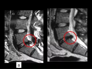
(a) This patient presented with acute low back pain and sciatica. He has the characteristic sideways list or tilt due to paravertebral muscle spasm. (b,c) MRI showing the disc prolapse at L5/S1. In the axial view (c), one can see that the disc protrusion encroaches on the intervertebral foramen and nerve root at that level.
Differential diagnosis 1. Inflammatory disorders, such as ankylosingspondylitis, cause severe and more generalized stiffness and typical x-ray changes. 2. Vertebraltumourscause constant pain; x-rays show bone destruction or a pathological fractures 3. Nerve tumoursmay cause sciatica but pain is continuous; CT or MRI may delineate the lesions.
:treatment Heat and analgesics soothe, and exercises strengthen muscles; but there are only three ways of treating the prolapse itself *rest *reduction * removal; *equally important is the rehabilitation afterwards.
Rest: -With an acute attack the patient should be kept in bed, with hips and knees slightly flexed and 10 kg traction to the pelvis. -An anti-inflammatory drug such as indomethacin is useful. -For mild attacks a spinal corset and reduced activity may suffice.
Reduce: Continuous bed-rest and traction for 2 weeks will reduce the herniation in more than 90 percent of cases. If the symptoms and signs have not improved significantly by then, an epidural injection of corticosteroid and local anesthetics may help. ** If conservative measures fail, discectomy is the treatment of choice.
Removal: The indications for operative removal of a disc are: (1) caudaequina compression syndrome which does not clear up within 6 hours of starting bed-rest and traction – this is an emergency (2) persistent pain and severely limited straight-leg raising after 2 weeks of conservative treatment (3) neurological deterioration while under conservative treatment; (4) frequently recurring attacks. Through a posterior approach between adjacent vertebral laminae(ligamentumflavum), the dural sac is retracted to one side and the bulging disc is exposed. The friable, partially shredded material is removed. This can be done by open operation or by endoscopic surgery (microdiscectomy).
Rehabilitation : After recovery from an acute disc rupture, or disc removal, the patient is taught isometric exercises and how to lie, sit, bend and lift with the least strain. Light work is resumed after a month and heavy work after 3 months. At that stage, if recovery is anything but total, the patient should be advised to avoid heavy lifting tasks altogether.
Recurrent pain after discectomy??? 1.infection 2.recurrent disc 3.wrong level