Comprehensive Overview of Male and Female Reproductive Anatomy and Physiology
This resource provides an in-depth look at the structures and functions of the male and female reproductive systems, including external genitalia, duct systems, and gamete production. It covers male anatomy such as the penis, testes, and accessory glands, explaining processes like erection and spermatogenesis. For females, it details the vagina, ovaries, and uterine cycle, including ovulation and oogenesis. The interrelationship of hormones and the mechanisms of reproduction are also discussed, offering a complete understanding of human reproductive physiology.

Comprehensive Overview of Male and Female Reproductive Anatomy and Physiology
E N D
Presentation Transcript
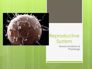
Reproductive System Honors Anatomy & Physiology
Structures & Their functions • External Genetialia: • Penis – copulatory organ • Shaft, glans (enlarged tip), prepuce (foreskin) • Erectile (spongy) tissue – connective tissue, smooth muscle, and vascular spaces • Erection – sexual arousal triggers parasympathetic release of NO causing arterioles to dilate, engorgement restricts drainage • Testes (gonads)-1.5” x 1” sperm & testosterone production • Scrotum – maintain testes temp 3oC lower than body temp (retractable) • Duct System (sympathetic control over ejaculation) • Epididymis –20 ft long, 20 days for sperm to become motile, may be stored for months (ejaculated by smooth muscle or phagocytized) • Ductus deferens – 18” long, thick smooth muscle • Ejaculatory duct – through the prostate and empties into urethra • Urethra – both urine & sperm (has priority!) • 3 regions: prostatic urethra, membranous, spongy
Semen • Semen – milky white, sticky secretion of sperm & secretions • 2-5mL per ejaculation • 20-150milliom sperm per mL • Catabolism of fructose provides ATP for flagella • Prostaglandins stimulate uterine peristalsis • Accessory Glands • Seminal vesicles – 60% of semen: yellow, viscous alkaline fluid w/fructose, ascorbic acid, prostaglandins enhancing sperm motility • Prostate Gland – 30% of semen: milky acidic fluid w/enzymes to activate sperm • Bulbourthral glands – pea sized glands secrete a thick clear mucus to neutralize acidic urine in urethra “pre-ejaculate”
Structure of the testis • Surrounded by 2 membranes: • Tunica vaginalis • Tunica albuginea • Divided into 250-300 lobules • Ea/w 1-4 seminiferous tubules (sperm production) • Surrounded by myoid cells – contract to squeeze sperm out of testis into rete testis
Spermatogenesis • Begins @ age 14 • Makes ~400million sperm/day • Spermatogonia – divide by mitosis • @puberty some become primary spermatocytes generating secondary spermatocytes • Producing non motile spermatotids
Female External Genitalia • Vagina – 3-4” birth canal & copulation organ • 3 layers: outer fibroelastic, smooth muscle, ridged mucosa • Acidic pH – antimicrobial but hostile to sperm • Hymen – vascular membrane in virgins • Mons pubis – fatty ridge • Labia: majora & minora– enclose the vestibule or entrance • Vestibular glands – secrete mucus • Clitoris – innervated erectile tissue hooded by prepuce
Ovaries • Produce Ova (eggs) & Sex hormones • Ovarian ligament anchor to uterus & suspensory ligament to pelvic wall • Ovarian follicles ea/contains oocytesurrounded by follicle cells • Primordial follicle – 1 layer cells surround oocyte • Primary follicle – 2 + layers of cells • Secondary follicle – fluid filled cavity appears • Graafian follicle – follicle bulges from ovary surface, mature oocyte • Ovulation – oocyte ejected from ovary • Corpus luteum– follicle degenerates into scars/pits
Oogenesis • Fetal period – oogonia divide by mitosis • (7 million) Primary oocytesin primary follicles begin meiosis I, and stall in prophase I (2 milion) • Puberty 250,000 oocytes remain • 1 selected every 28 days to complete meiosis I, first polar body undergoes apoptosis, secondary oocyteis suspended in metaphase II when ovulated • <500 released over 40 years • After fertilized, it completes meiosis II, creating a second polar body that deteriorates and 1 large ovum
21-40 days, avg 28 days) Ovarian Cycle • @puberty pituitary releases FSH & LH stimulating ovaries • Follicular Phase (days 1-14) • Menstration(days 1-5 ) • endometrium sheds • Low estrogen & progesterone • Proliferative Phase (day 6-14) • FSH & LH • follicle growth • estrogen (- feedback on release FSH & LH, + feedback on estrogen) • Endometrial lining thickens • Cervical mucus thins • Ovulation (day 14) • estrogen causes surge of LH & FSH, then drops • Ovary expels 2ooocyte w/corona radiata • Mittlschmerz– abdominal pain due to ovarian wall stretching • Luteal /Secretory Phase (days 14-28) • estrogen • progesterone (thickens endometrium) • Corpus luteum degenerates
Female Duct System • Uterine (fallopian) tubes • Receive ovulated oocyte • About 4” long • Fimbraie – ciliated finger like projections on distal end create current to capture oocytes • Infundibulum – funnel shaped structure • Smooth muscle (peristalysis) & mucosa ciliated lining • Uterous (womb)- nourish fertilized ovum • Inverted pear in size and shape • 3 regions: fundus, body, & cervix • 3 layers of wall: • Perimetrium– outermost layer • Myometrium – smooth muscle • Endometrium – 2 layers of mucosal lining (inner layer sheds)
Menopause • 20’s reproductive peak • 30’s ovarian function (quality of oocytes) declines, still ~100,000 oocytes left • By 50, only ~3 oocytes left • Estrogen production declines • Menstrual periods erratic & shorter, until ovulation & menstruation ceases • Menopause reached after 1 year w/o menstruation
Mammary Glands • Produce milk by modified sweat glands • Areola – ring of pigmented skin surrounding nipple • Ea gland/ 15-25 lobes consisting of lobules containing alveoli that produce milk • Milk passes into lactiferous ducts which open at nipple