Comprehensive Review of Foot and Toes Anatomy: Bones, Muscles, and Assessments
130 likes | 271 Vues
This thorough evaluation explores the anatomy of the foot, detailing its 26 bones, including the phalanges, metatarsals, and tarsals, and their respective sections: the rearfoot, midfoot, and forefoot. It covers vital articulations and ligaments, such as the subtalar and midtarsal joints, alongside the numerous intrinsic and extrinsic muscles influencing foot movement. Key points on history, injury mechanisms, inspection criteria, palpation, range of motion testing, ligamentous testing, and neurological assessments are addressed to ensure a comprehensive understanding for clinicians and students.

Comprehensive Review of Foot and Toes Anatomy: Bones, Muscles, and Assessments
E N D
Presentation Transcript
Evaluation-The Foot and Toes Ms. Bowman
Anatomy Review-Bones • 26 bones • Phalanges-toes; proximal, middle, and distal • Metatarsals-5; between phalanges and tarsals • Tarsals-calcaneus, talus, navicular, cuboid, 3 cuneiforms • Divided into 3 sections • Rearfoot-formed by the calcaneus and talus; provides stability and shock absorption during the initial stance phase of gait and serves as a lever arm for the Achilles tendon during plantarflexion • Midfoot-composed of the navicular, three cuneiforms, and cuboid; shock absorbing section of the foot • Forefoot and toes-formed by the 5 metatarsals and 14 phalanges; act as a lever during the preswing phase of gait
Anatomy Review-Articulations and Ligaments • Subtalar Joint-articulation of the calcaneus and talus • Midfoot- • midtarsal joint formed by the articulations of the tarsal bones • Plantar calcaneonavicular ligament (spring liegament) • Forefoot- • Tarsometatarsal joints-junction between the midfoot and forefoot • Intermetatarsal joints-proximal and distal joints • Metatarsophalangeal joints • Interphalangeal joints • Medial Ligaments • Deltoid ligament-composed of 4 ligaments • Posterior tibiotalar ligament • Tibiocalcaneal ligament • Anterior tibiotalar ligament • Tibionavicular ligament • Lateral Ligaments • Anterior talofibular ligament • Calcaneofibularligament • Posterior talofibular ligament
Anatomy Review-Muscles • There are many muscles that act on the foot. • Those that originate and insert in the foot are called intrinsic foot muscles. These directly influence the foot and toes. • Those that originate in the lower leg are called extrinsic foot muscles. These influence motion at the ankle and knee as well as the foot and toes. • If the muscle name begins with extensor, then the muscle’s primary function is extension. • If the muscle name begins with flexor, then the muscle’s primary function is flexion.
History • Location of p!-trauma to intrinsic structures or secondary to compensation for improper lower leg biomechanics • Metatarsal p!-p! that worsens over time can indicate stress fx; p! between the MTs possibly a result of nerve impingement • Great toe p!-localized p! to plantar surface can be indicative of sesamoidfx; p! with flexion or extension can be an indicator for turf toe • Onset and Mechanism of injury • Acute onset- can occur from trauma (fx, ligament sprain, muscle strain) • Insidious onset-result of overuse injuries; may be the result of playing surface, distance and duration of exercise, shoes
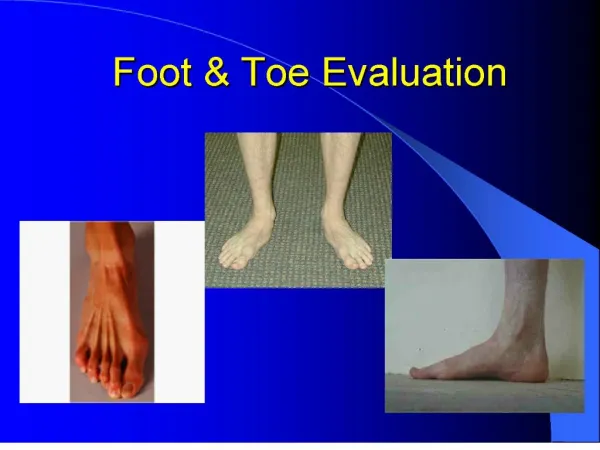
Inspection • Analyze gait • Look for • Gross deformity • Edema • Redness • Calluses and blisters (indicates improperly fitting shoes, poor mechanics, or underlying bony or soft tissue dysfunction) • Observe • Alignment of the toes • Toenail integrity (ingrown toenails, subungual hematoma) • Arches • Achilles • Calcaneus • Plantar surface of the foot
Palpation • When palpating, check for deformity, alignment, edema, crepitus, and pain • Medial Structures • 1st Phalange, 1st MT, 1st Cuneiform, Navicular, Talar head Spring Ligament, Calcaneus • Lateral Structures • 5th phalange, 5th MT, Cuboid, Peroneal tubercle, Calcaneus • Dorsal Structures • Rays, Cuneiforms, Navicular, Dome of Talus, Musculature • Plantar Structures • Plantar fascia, MT heads, sesamoid bones of great toe
ROM Testing • ROM should be measured with a goniometer • AROM, PROM, and RROM should be assessed as necessary • Goniometry measurements • Fulcrum-placed over joint • Stationary arm-placed over the proximal (non-moving part of body) • Movement arm-placed over the distal (moving part of body)
Ligamentous Testing • Valgus and Varus stress tests should be used to test the integrity of the MTP and IP joints • Metatarsal gilde test can be used to tests for the integrity of the ligaments connecting the MT • Midtarsal joint glide test can be used to test the integrity of the intertarsal ligaments
Neurological Assessment • Foot and toes are supplied by L4 and S2 nerve roots • Can be assessed by doing a lower quarter screen
Special Tests • Navicular drop test • Tinel’s sign • Long bone compression test • Pencil test