Download
1 / 25
290 likes | 899 Vues
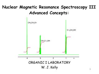
Nuclear Magnetic Resonance Spectroscopy III Advanced Concepts:. ORGANIC I LABORATORY W. J. Kelly. 1. Nuclear Magnetic Resonance Spectroscopy. 1 H NMR—Spin-Spin Splitting. Protons on carbon-carbon double bonds often give characteristic splitting patterns.

E N D
Nuclear Magnetic Resonance Spectroscopy III Advanced Concepts: ORGANIC I LABORATORY W. J. Kelly 1
Nuclear Magnetic Resonance Spectroscopy 1H NMR—Spin-Spin Splitting • Protons on carbon-carbon double bonds often give characteristic splitting patterns. • A disubstituted double bond can have two geminal protons, two cis protons, or two trans protons. • When these protons are different, each proton splits the NMR signal of the other so that each proton appears as a doublet. • The magnitude of the coupling constant J for these doublets depends on the arrangement of hydrogen atoms. 2
Nuclear Magnetic Resonance Spectroscopy 1H NMR—Spin-Spin Splitting 3
Nuclear Magnetic Resonance Spectroscopy Complex Splitting Patterns:Non-equivalent coupling 4
Nuclear Magnetic Resonance Spectroscopy Complex Splitting Patterns:Non-equivalent coupling thus far, we have concentrated on spin-spin coupling with only one other nonequivalent set of H atoms more complex splittings arise when a set of H atoms couples to more than one set H atoms a tree diagram shows that when Hb is adjacent to nonequivalent Ha on one side and Hc on the other, the resulting coupling gives rise to a doublet of doublets (Jab > Jbc) 5
Nuclear Magnetic Resonance Spectroscopy Complex Splitting Patterns:Non-equivalent coupling if Hc is a set of two equivalent H, then the observed splitting is a doublet of triplets 6
Nuclear Magnetic Resonance Spectroscopy 1H NMR—Spin-Spin Splitting:Non-equivalent coupling Now consider the spectrum of vinyl acetate. Since Hc and Hb are not equivalent to each other, we cannot merely add them together and use the n + 1 rule. When two sets of adjacent protons are different from each other and couple to a common set of protons with different J, (n protons on one adjacent carbon and m protons on the other), the number of peaks in an NMR signal = (n + 1)(m + 1). 7
Nuclear Magnetic Resonance Spectroscopy 1H NMR—Spin-Spin Splitting Splitting diagrams for the alkenyl protons in vinyl acetate are shown below. Note that each pattern is different in appearance because the magnitude of the coupling constants forming them is different.
Nuclear Magnetic Resonance Spectroscopy 1H NMR—OH Protons • Under usual conditions, an OH proton does not split the NMR signal of adjacent protons. • The signal due to an OH proton is not split by adjacent protons. 9
Nuclear Magnetic Resonance Spectroscopy 1H NMR—OH Protons • Ethanol (CH3CH2OH) has three different types of protons, so there are three signals in its NMR spectrum. • The Ha signal is split by the two Hb protons into three peaks (a triplet). • The Hb signal is split only by the three Ha protons into four peaks, a quartet. The adjacent OH proton does not split the signal due to Hb. • Hc is a singlet because OH protons are not split by adjacent protons. • Protons on electronegative atoms rapidly exchange between molecules in the presence of trace amounts of acid or base. Thus, the CH2 group of ethanol never “feels” the presence of the OH proton, because the OH proton is rapidly moving from one molecule to another. • This phenomenon usually occurs with NH and OH protons. 10
Nuclear Magnetic Resonance Spectroscopy 1H NMR—Cyclohexane Conformers • Recall that cyclohexane conformers interconvert by ring flipping. • Because the ring flipping is very rapid at room temperature, an NMR spectrum records an average of all conformers that interconvert. • Thus, even though each cyclohexane carbon has two different types of hydrogens—one axial and one equatorial—the two chair forms of cyclohexane rapidly interconvert them, and an NMR spectrum shows a single signal for the average environment that it “sees”. 11
Nuclear Magnetic Resonance Spectroscopy 1H NMR—Protons on Benzene Rings • Benzene has six equivalent deshielded protons and exhibits a single peak in its 1H NMR spectrum at 7.27 ppm. • Monosubstituted benzenes contain five protons that are no longer structurally equivalent will appear as a singlet when the substituent bonded to the ring is an sp3 hybridized carbon because of Magnetic Equivalence. That is they all have the same chemical shift. • If the atom or group bonded to the ring is other than an sp3 hybridized carbon, the nmr absorption region ismore complex 12
All H’s are equivalent - singlet Single Ha splits Hb into a doublet And Hb splits Ha into a doublet Single Ha splits Hb into a doublet And Hb splits Ha into a doublet
Nuclear Magnetic Resonance Spectroscopy 13C NMR 13C Spectra are easier to analyze than 1H spectra because the signals are not split. Each type of carbon atom appears as a single peak. 14
Nuclear Magnetic Resonance Spectroscopy 13C NMR • Provides a count of the different types of environments of carbon atoms in a molecule • 13C resonances are 0 to 220 ppm downfield from TMS (Figure 13-7) • Chemical shift affected by electronegativity of nearby atoms • O, N, halogen decrease electron density and shielding (“deshield”), moving signal downfield. • sp3 C signal is at 0 to 9; sp2 C: 110 to 220 • C(=O) at the low field, 160 to 220 15
Nuclear Magnetic Resonance Spectroscopy 13C NMR • The lack of splitting in a 13C spectrum is a consequence of the low natural abundance of 13C. • Recall that splitting occurs when two NMR active nuclei—like two protons—are close to each other. Because of the low natural abundance of 13C nuclei (1.1%), the chance of two 13C nuclei being bonded to each other is very small (0.01%), and so no carbon-carbon splitting is observed. • A 13C NMR signal can also be split by nearby protons. This 1H-13C splitting is usually eliminated from the spectrum by using an instrumental technique that decouples the proton-carbon interactions, so that every peak in a 13C NMR spectrum appears as a singlet. • The two features of a 13C NMR spectrum that provide the most structural information are the number of signals observed and the chemical shifts of those signals. 16
Nuclear Magnetic Resonance Spectroscopy 13C NMR—Number of Signals • The number of signals in a 13C spectrum gives the number of different types of carbon atoms in a molecule. • Because 13C NMR signals are not split, the number of signals equals the number of lines in the 13C spectrum. • In contrast to the 1H NMR situation, peak intensity is not proportional to the number of absorbing carbons, so 13C NMR signals are not integrated. 17
Nuclear Magnetic Resonance Spectroscopy 13C NMR—Position of Signals • In contrast to the small range of chemical shifts in 1H NMR (1-10 ppm usually), 13C NMR absorptions occur over a much broader range (0-220 ppm). • The chemical shifts of carbon atoms in 13C NMR depend on the same effects as the chemical shifts of protons in 1H NMR. 18
Nuclear Magnetic Resonance Spectroscopy 13C NMR—Number of Signals 24
Nuclear Magnetic Resonance Spectroscopy 13C NMR—Number of Signals 25