Ligaments and Tendons Digital Laboratory
180 likes | 414 Vues
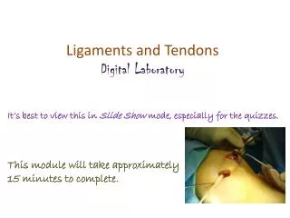
Ligaments and Tendons Digital Laboratory. It’s best to view this in Slide Show mode, especially for the quizzes. This module will take approximately 15 minutes to complete. After completing this exercise, you should be able to:

Ligaments and Tendons Digital Laboratory
E N D
Presentation Transcript
Ligaments and Tendons Digital Laboratory It’s best to view this in Slide Show mode, especially for the quizzes. This module will take approximately 15 minutes to complete.
After completing this exercise, you should be able to: • Distinguish, at the light microscope level, each of the following components of connective tissue: • Review components of connective tissue seen in H&E sections • Fibroblasts • Collagen fibers • Review types of generic connective tissue • Loose (areolar) connective tissue • Dense irregular connective tissue • Dense regular connective tissue • Distinguish, at the electron microscope level, each of the following components of connective tissue: • Review components of connective tissue • Fibroblast • Collagen fibers The only new thing here is identifying dense regular connective tissue, and we briefly introduced that in Fundamentals.
CLASSIFICATION OF CONNECTIVE TISSUE Connective tissue can be classified into generic and specialized : Generic connective tissues --Loose (aka areolar, includes reticular, elastic, maybe mesenchyme) --Dense irregular (aka dense irregular) --Dense regular (aka dense regular) This is a review slide from the digital lab on generic connective tissues. We are revisiting dense regular in this module. We care about these today Specialized connective tissues --adipose --cartilage (hyaline, elastic, fibrous) --bone --blood These will be covered later.
CLASSIFICATION OF CONNECTIVE TISSUE Fibroblast nuclei Collagen fibers: Recall that generic connective tissues are characterized in H&E sections by collagen fibers and fibroblast nuclei.
CLASSIFICATION OF CONNECTIVE TISSUE In loose connective tissue, the collagen fibers are spread out, whereas in dense irregular connective tissue, the fibers are more closely packed together. In both cases, the collagen fibers are oriented in all directions to provide 3-dimensional strength. Both contain numerous blood vessels. In this image, recall loose connective tissue (black outline) and dense irregular connective tissue (blue outline).
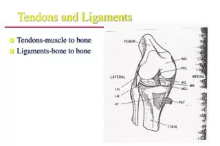
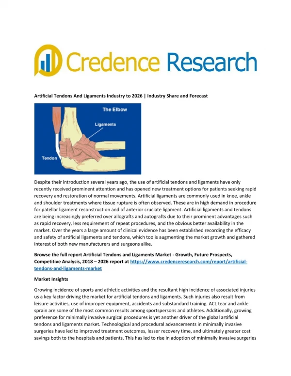
LIGAMENTS AND TENDONS Ligaments and tendons are similar structures: ligaments connect bone to bone tendons connect muscle to bone Histologically, both are dense regular connective tissue. We only have a slide of a tendon, so that’s what you’ll see here.
DENSE REGULAR CONNECTIVE TISSUE Fibroblast nuclei Collagen fibers (basically everywhere) Dense regular connective tissue appearance – no space, many thick (type I) collagen fibers packed so tightly, it’s hard to see individual fibers, so it is dense collagen fibers oriented in one direction, this is difficult to appreciate because the individual collagen fibers are hard to see, but look how the fibroblast nuclei have to orient and narrow to accommodate, so it is regular function – strong in one direction (e.g. tendon, ligament), but usually very poorly vascularized, so it takes a long time to heal
DENSE REGULAR CONNECTIVE TISSUE Video of tendon showing dense regular connective tissue – SL27 • Link to SL 027 • Be able to identify: • Dense regular connective tissue
DENSE REGULAR CONNECTIVE TISSUE In this electron micrograph from a tendon, note obvious fibroblast with rough ER (4) and Golgi (3). Processes of fibroblasts are indicated at 6. Note collagen fibers (7) are all oriented in the same direction.
LIGAMENTS AND TENDONS On a slide, you can’t tell whether you are looking at a ligament or tendon, so better to say “dense regular connective tissue” (we’ll be asking for tissue type anyway). In the gross lab, you’ll want to give these structures specific names (e.g. patellar ligament).
The next set of slides is a quiz for this module. You should review the structures covered in this module, and try to visualize each of these in light and electron micrographs. • Distinguish, at the light microscope level, each of the following components of connective tissue: • Review components of connective tissue seen in H&E sections • Fibroblasts • Collagen fibers • Review types of generic connective tissue • Loose (areolar) connective tissue • Dense irregular connective tissue • Dense regular connective tissue • Distinguish, at the electron microscope level, each of the following components of connective tissue: • Review components of connective tissue • Fibroblast • Collagen fibers
Final quiz Self-check: Identify the tissue. (advance slide for answers) Dense irregular connective tissue
Final quiz Self-check: Identify the outlined tissue. (advance slide for answers) Hyaline cartilage
Final quiz Self-check: Identify the outlined tissue. (advance slide for answers) Smooth muscle Nuclei a little too plump, and too many nuclei to be connective tissue
Final quiz Self-check: Identify the outlined structure. (advance slide for answers) Peripheral nerve
Final quiz Self-check: Identify the tissue. (advance slide for answers) Dense regular connective tissue
Final quiz Self-check: Identify the outlined tissue. (advance slide for answers) Loose connective tissue
Final quiz Self-check: Identify the outlined structure. (advance slide for answers) Peripheral nerve