Download
1 / 74
750 likes | 1.24k Vues
Evaluation and Management of Acute Stroke. Mark I. Harris, M.D. Comprehensive Neurology Specialists Clinical Assistant Professor Emory Department of Family Medicine. Guidelines for the Early Management of Adults with Ischemic Stroke From the AHA/ASA Stroke Council.

E N D
Evaluation and Management of Acute Stroke Mark I. Harris, M.D. Comprehensive Neurology Specialists Clinical Assistant Professor Emory Department of Family Medicine
Guidelines for the Early Management of Adults with Ischemic StrokeFrom the AHA/ASA Stroke Council Harold P. Adams, Chair; Gregory del Zoppo, Vice-Chair, Mark J. Alberts, Deepak L Bhatt, Lawrence Brass, Anthony Furlan, Robert L. Grubb, Randy Higashida, Edward C. Jauch, Chelsea Kidwell, Patrick D. Lyden, Lewis B. Morgenstern, Adnan I. Qureshi, Robert H. Rosenwasser, Philip A. Scott, Eelco FM Wijdicks Stroke 2007; 38(5): 1655-1711
Introduction Since the 2003 AHA Stroke Council guidelines, evidence has been published to further refine the approach to the patient with acute ischemic stroke. In some cases, where supportive evidence from clinical trials was not available, the panel made a specific recommendation on the basis of pathophysiology and expert practice experience.
Example Case • A 47 year old female presents to the emergency department with onset 6-8 hours ago of • Right arm weakness: mild • Complains of dizziness • Exam: 190/ 110 • Mild drift right arm • 4/5 arm and leg • Speech normal
Example Case • CT brain: chronic white matter changes • BP: 190/110 • Treated w/ Procardia and clonidine
Example Case • 30 minutes later • 160/ 70 • Dense right hemi-paresis • Aphasia • MRI large left MCA diffusion defect
STROKE • 3rd leading cause of death • 167,400 deaths in 1999 • U.S.A.: 1 stroke / minute 1 death every 3.4 min • Leading cause of adult disability • Risk greatest in elderly, men, & blacks • Signs, symptoms or risk factors unknown to most people • Death rate 10% higher in the southeastern states (stroke belt) N Eng J Med. 1995;333:1392-4000
Modifiable Stroke Risk Factors • Behaviors • Cigarette smoking • Alcohol abuse • Physical inactivity Medical Conditions • Hypertension • Cardiac disease • Atrial fibrillation • Hyperlipidemia • Diabetes mellitus • Carotid stenosis • Prior TIA or stroke • Elevated homocysteine Sacco RL, et al. Stroke 1997;28:1507-1517. Pancioli AM, et al. JAMA 1998;279:1288-1292.
Stroke Risk Factors • AGE: RISK DOUBLES EVERY 10 YRS AFTER 55Y/O • SEX: 30% > RISK FOR MEN • RACE: AFRICAN-AMERICANS >60% HIGHER RISK OF DEATH AND DISABILITY FROM STROKE
Potential Stroke Risk ReductionAHA Guidelines Factor Risk reduction with treatment Hypertension 30% - 40% Smoking 50% w/in 1 yr, baseline >5 yrs Diabetes 44% reduction in HTN diabetics with tight blood pressure control Hyperlipidemia 20-30% with statins in patients with CAD Atrial fibrillation (non-valvular) 68% (warfarin) 21% (aspirin) Adapted from Goldstein, et al. Circulation 2001;103:163-182.
COMMONLY USED DEFINITIONS • TIA: ISCHEMIC EVENT LASTING MINUTES WITHOUT RESIDUAL NEUROLOGIC DEFICIT • COMPLETED STROKE: ISCHEMIC EVENT WITH PERSISTENT DEFICIT • STROKE in EVOLUTION: WORSENING of DEFICIT OVER TIME WHICH SUGGESTS INCREASING AREA OF BRAIN ISCHEMIA
Inclusion criteria: Objective:Outcome measures: Total events: TIA by ED physicians Short-term risk of strokeafter ED diagnosisRisk of stroke and otherevents during the 90 daysafter index TIA 25.1% 30.0% Short-term Prognosis after ER Diagnosis of TIA Outcome events 25.0% 20.0% 15.0% 12.7% 10.5% 10.0% Within90 days 5.0% 2.6% 2.6% 5.3% Within48 hr 0.0% Stroke CV event Death Recurrent TIA Johnston SC, et al. JAMA 2000;284:2901-2906.
Slightdisabilities (mRS 0-2) Follow-up after 90 days Prognosis of Ischemic StrokeGerman Stroke Data Bank Moderate disabilities (mRS 3) Severe disabilities (mRS 4-5) 14.70% 18.60% Deceased n = 5,017 9.40% 57.20% Grau AJ, et al. Stroke 2001;32:2559-2566.
Deaths from stroke or MI among patients with a first cerebral infarction (n = 764) Mortality Following Stroke 30 27.0% 25 20 Percentage 15 10.1% 10 5 0 Stroke Deaths MI Deaths • In data acquired over an 18-year observation period, for those with an initial stroke, the risk of death from stroke is more than 2.5 times the risk of death from MI Petty GW, et al. Neurology 1998;50:208-216.
A Stroke Is Not a “Heart Attack of the Brain” COMPARED TO MI • Causes of stroke are more heterogeneous • Hemorrhage can be a devastating complication of ischemic stroke • Strokes occur in older patients • Strokes have a higher risk of recurrence • MI patients are at > risk for a second MI
Stroke Subtypes Lacunar 19% Thrombo-embolic 6% SAH 13% Cardio-embolic 14% Ischemic 71% Hemorrhagic 26% ICH 13% Unknown 32% Other 3% NINDS Stroke Data Bank: Foulkes, et al. Stroke 1988;19:547.
WARNING SIGNS OF STROKE SUDDEN ONSET OF SYMPTOMS: • BLURRED OR DECREASED VISION • NUMBNESS, WEAKNESS, PARALYSIS • DYSARTHRIA • DYSPHASIA • DIZZINESS • ATAXIA • ABRUPT ONSET OF HEADACHE
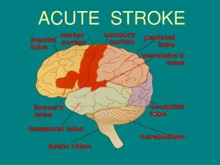
Carotid territory Amaurosis fugax Dysphasia Hemiparesis Hemi-sensory loss Vertebrobasilar Hemianopia Quadraparesis Cranial N dysfunction Cerebellar syndrome Crossed deficit Loss of consciousness Localization
Carotid bruit • If present, 60-70% of patients have an internal carotid stenosis BUT • False negative ex. very tight (>95%) stenosis • False positive ex. external carotid stenosis or transmission from aortic valve
Diagnosis of Carotid Stenosis • Carotid Duplex Ultrasound • Sensitivity of 94 - 100% for > 50% stenosis • May overdiagnose occlusion • Non-invasive
Diagnosis of Carotid Stenosis • Magnetic Resonance Angiography • Similar sensitivity to carotid ultrasound • Overestimates degree of stenosis • Gives information about vertebrobasilar system • Accuracy of 62% in detecting intracranial pathology • Cost and claustrophobia
Diagnosis of Carotid Stenosis • Cerebral Angiography • Gold standard for diagnosis • Invasive, with risk of stroke 1% • Patients with positive ultrasound • Patients with occlusion on US • First test if intracranial pathology suspected
Surgical Treatment Carotid endarterectomy (CEA) • Consider CEA if life expectancy > 2yrs, • Treating center complication rate < 5% • Symptomatic carotid stenosis • 70-99% • 50-69%- NNT 15 over 5 yrs • Asymptomatic carotid stenosis • 70-99% only VA/DoD clinical practice guideline for the management of stroke rehabilitation. www.qmo.amedd.army.mil. Oct 2002. Accessed 9 Jan 2004.
Changes from 2003 Guidelines • Pre-hospital care • Stroke Centers • Detailed recommendations on supportive care • Multimodal Imaging • Recommendation to RESTART anti-hypertensive's at 24 hours after onset. • Recommend re: thrombolysis eligibility & delivery • Recommendations AGAINST clopidogrel acutely • Recommendations AGAINST using experimental strategies (hyperbaric O2, Merci device, glycoprotein IIb-IIIa antagonists, drug-induced hypertension, combination reperfusion strategies) Adams et al. Stroke 2003; 34:1056-1083
AHA Classes and Levels of Evidence • Class I Agreement the treatment is useful and effective • Class II Conflicting evidence about the usefulness/efficacy of a treatment. • Class IIa Weight of evidence is in favor • Class IIb Efficacy is less well established • Class III Evidence and/or general agreement that the treatment is not useful/effective and in some cases may be harmful. • Levels of Evidence A: Data derived from multiple randomized trials. B: Data derived from a single randomized trial or nonrandomized studies. C: Consensus opinion of experts.
Pre-hospital • New: To increase the number of patients who are treated, educational programs for physicians, hospital personnel, and EMS personnel also are recommended (Class I, Level B) • New : Brief assessment by EMS personnel are recommended (Class I, Level B) • New :The use of a stroke algorithm is encouraged (Class I, Level B)
Pre-hospital • New: Recommends that EMS personnel begin the initial management of stroke in the field (Class I, Level B) • New : The development of stroke protocols to be used by EMS personnel is strongly encouraged. Patients should be transported rapidly for evaluation and treatment to the closest institution that provides emergency stroke care (Class I, Level B) • New: Telemedicine can be an effective method to extend acute stroke care to rural areas. (Class IIb, Level B)
Emergency Diagnosis and Management:Class I recommendations • The goal is to complete an evaluation and decide on treatment within 60 minutes. (Class I, Level B) • Stroke rating scale is recommended (e.g., NIH Stroke Scale) (Class I, Level B)
Emergency Diagnosis and Management • An ECG is recommended (Class I, Level B) • Basic blood tests are recommended (Class I, Level B) • Patients with clinical evidence of acute cardiac or pulmonary disease may warrant a CXR (Class I, Level B)
Brain and Vascular Imaging : Class I recommendations • Imaging of the brain is recommended before initiating specific therapy (Class I, Level A) • In most instances, CT will provide the information to make decisions about emergency management (Class I, Level A) • New: The imaging study should be interpreted by a physician with expertise in reading brain CT and MRI(Class I, Level C)
Brain and Vascular Imaging: Class II Recommendations • Other than hemorrhage, no specific CT finding should preclude use of IV r-tPA within 3 hours of stroke onset (Class II-b, Level A) • Vascular imaging is required before intra-arterial or endovascular interventions (Class II-a, Level B)
General Supportive Care: Class I Recommendations • Airway support and ventilatory assistance are recommended for the treatment of acute stroke (Class I, Level C) • Hypoxic patients with stroke should receive supplemental oxygen (Class I, Level C) • Fever should be treated and antipyretic medications should administered to lower temperature in febrile patients (Class I, Level C)
General Supportive Care • Cardiac monitoring to screen for atrial fibrillation and other potentially serious cardiac arrhythmias should be performed during the first 24 hours after onset of ischemic stroke (Class I Level B) • The management of arterial hypertension remains controversial. It is generally agreed that a cautious approach to the treatment of arterial hypertension should be recommended (Class I, Level C)
General Supportive Care • Patients who have elevated blood pressure and are otherwise eligible for treatment of r-tPA may have their blood pressure lowered so that their systolic is< 185 mm Hg and their diastolic blood pressure is < 110 mm Hg (Class I, Level B) • New: Until other data become available, consensus exists that the previously described blood pressure recommendations should be followed in patients undergoing other acute interventions to re-canalize occluded vessels, including intra-arterial thrombolysis (Class I, Level C)
Elevated in most patients with acute strokeBP drops spontaneously in the first days after strokeBlood flow in the critical penumbra passively dependent on the mean arterial pressure Blood pressure
Blood pressure • No controlled studies guiding BP management • Elevated BP 200/110 may be tolerated in the acute phase of ischemic stroke without intervention • BP may be lowered for cardiac conditions • Max. BP for thrombolytic therapy 180mmHg • Avoid / treat hypotension or drastic reduction in BP
General Supportive Care • New : Patients with markedly elevated blood pressure may have their blood pressure lowered. A reasonable goal is ~ 15% during the first 24 hours after onset of stroke. Medications should be withheld unless SPB > 220 or MAP >120 (Class I, Level C) • The cause of arterial hypotension in the setting of acute stroke should be sought (Class I, Level C) • New: Hypoglycemia should be treated in patients with acute ischemic stroke (Class I, Level C). Marked elevations in blood glucose should be avoided.
General Supportive Care: Class II Recommendations • New: There is no data to guide selection of medications for the lowering of blood pressure in the setting of acute ischemic stroke. The recommended medications and doses are based on general consensus (Class II-a, Level C) • New:For patients with preexisting hypertension evidence indicates antihypertensive therapy medications should be restarted at ~ 24 hours (Class II-a, Level B) • New: Persistent hyperglycemia (>140 mg/dL) during the first 24 hours after stroke is associated with poor outcomes (Class II-a, Level C)
General Supportive Care: Class III Recommendations • Non-hypoxic patients with acute ischemic stroke do not need supplemental oxygen therapy (Class III, Level B • New: Data on hyperbaric oxygen are inconclusive, and some data imply that the intervention may be harmful (Class III, Level B) • Despite the efficacy of hypothermia for improving neurological outcomes after cardiac arrest, the utility of induced hypothermia for the treatment of patients with acute ischemic stroke is not established (Class III, Level B)
Body temperature • Fever negatively influences neurological outcome • Experimentally, fever increases infarct size • Many patients with acute stroke develop a febrile infection • No adequately sized trials guiding temperature management • Consider treating fever & its cause when temperature > 37.5°C
Intravenous Thrombolysis: Class I Recommendations • IV r-tPA (0.9 mg./kg, maximum dose 90 mg) is recommended for selected patients who may be treated within 3 hours of onset of ischemic stroke (Class I, Level A) • New: Besides bleeding complications, physicians should be aware of the potential side effect of angio-edema that may cause partial airway obstruction (Class I, Level C)
DECIDING TO USE THROMBOLYTICS • UP TO 20% OF PATIENTS INCORRECTLY DIAGNOSED w/ STROKE • EXPERTISE IN STROKE MANAGEMENT NEEDED • EXCLUSION: • CT SIGNS OF HEMORRHAGE OR INFARCTION • UNDETERMINED TIME OF ONSET • UNCONTROLLED HYPERTENSION • RAPIDLY RESOLVING NEURO DEFICITS • UNRESOLVING NEURO DEFICIT LASTING > 90 MIN, LESS THAN 3% ARE TIA’s
Intravenous Thrombolysis: Class II Recommendations • A patient whose BP can be safely lowered with antihypertensive agents may be eligible for treatment. (Class II-a, Level B) • New: A patient with a seizure at the time of onset may be eligible for treatment. (Class II-a, Level C)
Intravenous Thrombolysis: Class III Recommendations • IV streptokinase for treatment of stroke IS NOT recommended. (Class III, Level A) • New: Use of IV fibrinolytics other than r-tPA (reteplase, tenecteplase, etc) outside a clinical trial in NOT recommended. (Class III, Level C)
Intra-arterial Thrombolysis: Class I Recommendations • Intra-arterial thrombolysis is an option for major stroke if administered within 6 hours of onset. ( Class I, Level B) • Intra-arterial thrombolysis should only be attempted at experienced stroke centers (Class I, Level C)