Download
1 / 42
420 likes | 673 Vues
Evaluation and Management of Acute Renal Failure. 范揚智醫師 95/10/12. 腎臟的功能. 調節水份 調節酸鹼 調節電解質 尿液的形成 內分泌器官 – Vitamin D, erythropoietin, RAS. Anuria: urine < 100ml/day Oliguria: urine <400-500 mL/d Azotemia: Increase Cr, BUN May be prerenal, renal, postrenal

E N D
Evaluation and Management of Acute Renal Failure 范揚智醫師 95/10/12
腎臟的功能 調節水份 調節酸鹼 調節電解質 尿液的形成 內分泌器官– Vitamin D, erythropoietin, RAS
Anuria: urine < 100ml/day Oliguria: urine <400-500 mL/d Azotemia: Increase Cr, BUN • May be prerenal, renal, postrenal • Does not require any clinical findings Chronic Kidney Disease: graduated loss of renal function, National kidney fundation: stage 1~5 ESRD = Loss of kidney function >3 months
Blood Urea Nitrogen (BUN) • Catabolism of aminoacids generates NH3 NH2 2 NH3 + CO2 = C = 0 + H2O NH2 • Urea Mol wt : 60; BUN Mol wt. : 28 • Normal BUN 10-20 mg/dl • After filtration › 50% is reabsorbed by the tubule • BUN level is related to: Renal function, protein intake, liver function, GI bleeding, steroid, hyper catabolic states
Creatinine • Formed at a constant rate by dehydration of muscle creatine • Normally 1–2% of muscle creatine is broken into creatinine • Mol. Wt. 113 • Creatinine is freely filtered by the glomerulii and is not reabsorbed 10–15% is secreted into proximal tubule
GFR Estimation by Plasma Creatinine • Cockcroft and Gault Formula* Calculated creatinine clearance = (140–age) x wt (kg) 72 X serum creatinine(mg/dl) For females, subtract 15% (or multiply by 0.85); for paraplegics multiply by 0.8, for quadriplegics, multiply by 0.6 *Applicable only when patient is in a steady state, not edematous and not obese
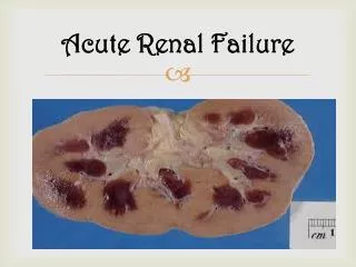
Acute Renal Failure Definition: • Rapid (hours to weeks) decline in glomerular filtration rate and retention of waste products • It is a clinical syndrome cause by many renal or extrarenal diseases • Lack a uniform definition • Cr > 1.5x, urine output <0.5ml/kg/hr • Cr increase ≥ 1.0 mg/dl/2d
The Second International Consensus Conference of the Acute Dialysis Quality Initiative (ADQI) Group
The facts you need to know about ARF • Acute renal failure may reversible and should look for the causes to management • Incidence: - 2-5% of hospitalized patients(55% iatrogenic) - 7-23% of ICU patients - 20-60% require dialysis; of those who survive initial dialysis, <25% require long-term dialysis
The facts you need to know about ARF Motality: • Liano(1996) reported mortality rate of 60% for patient with ATN, 35% for acute on chronic renal failure, 27% for obstructive ARF and 26% for renal disorder other than ATN. • Knaus(1986)50% for combination of acute renal and respiratory failure towards 100% with 5 system failure • In community-acquired ARF with mostly prerenal and postrenal causes and the prognosis is better. • Rates not significantly decreased over past 50 years despite advances in dialysis and critical care (increased patient age and co morbid illnesses)
Symptoms and Signs of Renal Failure • Retention of nitrogenous waste products • Nausea, vomiting, diarrhea, hiccup, foul taste, dry crusted mouth, itching, • Drowsiness, clouding of consciousness, neuropathy, pericarditis, GI bleeding, • Coma • Retention of salt and water • Pulmonary edema, peripheral edema, ascites, pleural effusion
Symptoms and Signs of Renal Failure • Retention of potassium • Weakness, lassitude, paralysis, EKG changes with tenting T waves, widening of QRS complex, increased PR interval, sine wave pattern, cardiac arrest, VT • Retention of acid • Kussmaul respiration, hyperreflexia, hypotension
A: Cr, BUN B: H, P, K C: NaCl
Classification of ARF Acute Renal Failure Pre-renal Intrinsic Post-renal Glomerular Interstitial Tubular Vascular
Pre-renal ARF • Accounts for 60-70% of cases of ARF • Represents physiologic response to mild-moderate renal hypoperfusion • Renal parenchymal tissue is not damaged therefore rapidly reversible upon restoration of RBF and glomerular filtration pressure • Elderly and those with pre-existing renal disease at increased risk
Pre-renal ARF I. Hypovolemia • A. Hemorrhage, burns, dehydration • B. GI fluid loss: vomiting, surgical drainage, diarrhea • C. Renal fluid loss: diuretics, osmotic diuresis (e.g., diabetes mellitus), hypoadrenalism • D. Sequestration in extravascular space: pancreatitis, peritonitis, trauma, burns, severe hypoalbuminemia II. Low cardiac output • A. Diseases of myocardium, valves, and pericardium; arrhythmias; tamponade • B. Other: pulmonary hypertension, massive pulmonary embolus, positive pressure mechanical ventilation
Pre-renal ARF III. Altered renal systemic vascular resistance ratio • A. Systemic vasodilatation: sepsis, antihypertensives, afterload reducers, anesthesia, anaphylaxis • B. Renal vasoconstriction: hypercalcemia, norepinephrine, epinephrine, cyclosporine, tacrolimus, amphotericin B • C. Cirrhosis with ascites (hepatorenal syndrome) IV. Renal hypoperfusion with impairment of renal autoregulatory responses • Cyclooxygenase inhibitors, ACEI V. Hyperviscosity syndrome (rare) • Multiple myeloma, macroglobulinemia, polycythemia
Intrinsic Renal Causes • Accounts for 25-40% of cases of ARF • Types: • Acute glomerulonephritis <5% • Interstitial nephritis 10% • Intrarenal vascular disease <5% • ATN 85%
Intrinsic Renal Causes I. Renovascular obstruction (bilateral or unilateral in the setting of one functioning kidney) • A. Renal artery obstruction: atherosclerotic plaque, thrombosis, embolism, dissecting aneurysm, vasculitis • B. Renal vein obstruction: thrombosis, compression II. Disease of glomeruli or renal microvasculature • A. Glomerulonephritis and vasculitis • B. Hemolytic uremic syndrome, thrombotic thrombocytopenic purpura, disseminated intravascular coagulation, toxemia of pregnancy, accelerated hypertension, radiation nephritis, systemic lupus erythematosus, scleroderma
Intrinsic Renal Causes III. Acute tubular necrosis • A. Ischemia(60%): as for prerenal ARF (hypovolemia, low cardiac output, renal vasoconstriction, systemic vasodilatation), obstetric complications (abruptio placentae, postpartum hemorrhage) • B. Toxins(40%) 1. Exogenous: radiocontrast, cyclosporine, antibiotics (e.g., aminoglycosides), chemotherapy (e.g., cisplatin), organic solvents (e.g., ethylene glycol), acetaminophen, illegal abortifacients 2. Endogenous: rhabdomyolysis, hemolysis, uric acid, oxalate, plasma cell dyscrasia (e.g., myeloma)
Intrinsic Renal Causes IV. Interstitial nephritis • A. Allergic: antibiotics (e.g., -lactams, sulfonamides, trimethoprim, rifampicin), nonsteroidal anti-inflammatory agents, diuretics, captopril • B. Infection: bacterial (e.g., acute pyelonephritis, leptospirosis), viral (e.g., cytomegalovirus), fungal (e.g., candidiasis) • C. Infiltration: lymphoma, leukemia, sarcoidosis • D. Idiopathic V. Intratubular deposition and obstruction • Myeloma proteins, uric acid, oxalate, acyclovir, methotrexate, sulphonamides VI. Renal allograft rejection
Post-renal Causes of ARF • Account for 5% of cases of ARF • ARF occurs when both urinary outflow tracts are obstructed or when one tract is obstructed in a patient with a single functional kidney
Post-renal Causes of ARF I. Ureteric • Calculi, blood clot, sloughed papillae, cancer, external compression (e.g., retroperitoneal fibrosis) II. Bladder neck • Neurogenic bladder, prostatic hypertrophy, calculi, cancer, blood clot III. Urethra • Stricture, congenital valve, phimosis
Investigations ARF: Focused History • Nausea? Vomiting? Diarrhea? • Hx of heart disease, liver disease, previous renal disease, kidney stones, BPH? • Any recent illnesses? • Any edema, change in urination? • Any new medications? • Any recent radiology studies? • Rashes?
Investigations Physical Examination: • Infection sign ? Blood pressure, vital sign • Volume Status • Mucus membranes, orthostatics, skin turgor, Edema • Cardiovascular • JVD, rubs • Pulmonary • Decreased breath sounds • Rales • Abd and back: bladder distension, masses, ascites, CVA tenderness, large prostate • Rash (Allergic interstitial nephritis)
Investigations • Blood • CBC-DC • Electrolyte, Ca, Mg, P • Urea, Creatinine • Others: LDH, Alb, CRP… • Urine • Urine sodium, Cr • Urine osmolality • Urinalysis • Renal echo
FeNa = (urine Na x plasma Cr) (plasma Na x urine Cr) FeNa <1% 1. PRERENAL • Urine Na < 20. Functioning tubules reabsorb lots of filtered Na 2. ATN (unusual) • Postischemic dz: most of UOP comes from few normal nephrons, which handle Na appropriately • ATN + chronic prerenal dz (cirrhosis, CHF) 3. Glomerular or vascular injury • Despite glomerular or vascular injury, pt may still have well-preserved tubular function and be able to concentrate Na
More FeNa FeNa 1%-2% • Prerenal-sometimes • ATN-sometimes • AIN-higher FeNa due to tubular damage FeNa >2% • ATN Damaged tubules can't reabsorb Na Calculating FeNa after pt has gotten Lasix. 1. Fractional Excretion of Lithium (endogenous) (<7% in prerenal ) 2. Fractional Excretion of Uric Acid (<7% in prerenal )
Is the renal failure really acute? Factors suggesting chronicity • Duration of symptoms for months • Absence of acute illness in the face of very high urea and creatinine • Anaemia of chronic disorders • Bone disease (renal osteodystrophy) • Sexual dysfunction • Skin disorders, pruritus • Neurological complications • Small kidneys on renal imaging
Management • Prevention • Etiology treatment • Prevention additional injury • Treatment of complication • Conservative measurement • Renal replacement therapy
Prevention • Identification of high-risk patients for pharmacologic agents-induced nephrotoxicity • iodinated radiocontrast medium, NSAIDs • Aggressive surveillance for nephrotoxin-induced renal dysfunction • cisplatin, amphotericin B, aminoglycoside • Use of volume expansion in selected clinical settings • Hyperpigmenturia: hemoglobinuria, myoglobinuria • Crystaluria: uric acid, acyclovir, methotrexate, sulfonamides • Minimalization of catheters use to avoid nosocomial sepsis
Etiology Treatment • Correct postrenal factor • Correct prerenal factor • Treat underlying sepsis • Stop nephrotoxic drugs
Conservative Measurement • Fluid balance • Careful monitoring of I/O and body weight • Fluid restriction • (usually less than 1 L/day in oliguric ARF) • Total intake < urine output +extrarenal losses • Electrolytes and acid -base balance • hyperkalemia • hyponatremia • Keep serum bicarbonate >15 • hyperphosphatemia • Treat hypocalcemia only if symptomatic
Guide of Volume Expansion • CVP 8-14 cm H2O (5-2 rule) • PAWP 12-16 mmHg (7-3 rule) • Urine output 0.5-1.0ml/kg/hour • Weighing the patient daily • Insensible water loss from the skin and respiratory tract (500 ml/day)
Dietary modification • total caloric intake– 35~ 50 kcal/kg/day to avoid catabolism Salt restriction– 2~4 g/day Potassium intake– 40 meq/day Phosphorus intake– 800 mg/day
Conservative Measurement • Uremia-nutrition • Restriction protein is not necessary in ARF, maintain caloric intake • Carbohydrate ≥ 100gm/day to minimize ketosis and protein catabolism • Drug • Review all medication, Stop magnesium-containing medication • Adjusted dosage for renal failure, Readjust with improvement of GFR
Indication of Dialysis Absolute indication • CCr 5 ml/min or serum Cr 10.0 mg/dl Relative indication • CCr 10 ml/min or serum Cr 8.0 mg/dl • With accompanied symptoms or signs = CHF/Pulmonary edema = Uremic pericarditis= Bleeding tendency = Neurologic symptoms= Drug-resistant hyper-K = Drug-resistant metabolic acidosis = Drug-resistant nausea/vomiting= Others
Take Home Points • Features of the history and physical examination in addition to relevant lab and radiologic investigations help to determine the most likely cause(s) of ARF in a given patient
Take Home Points • Management of a patient with ARF involves: • Treating potentially life-threatening complications • Reversing pre-renal and post-renal causes • Minimizing further hemodynamic and toxic insults to the kidney • Admission and appropriate consultation • Lack of evidence for converting oliguric to non-oliguric ARF