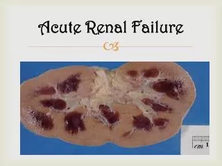
ACUTE RENAL FAILURE
ACUTE RENAL FAILURE. Fadi Jehad Zaben RN MSN IMET 2000, Ramallah. What is Acute renal failure?. Acute renal failure is a syndrome of varying causation that results in a sudden decline in renal function.


ACUTE RENAL FAILURE
E N D
Presentation Transcript
ACUTE RENAL FAILURE Fadi Jehad Zaben RN MSN IMET 2000, Ramallah
What is Acute renal failure? • Acute renal failure is a syndrome of varying causation that results in a sudden decline in renal function. • Acute Renal Failure (ARF) is characterized by rapid deterioration of renal function leading to accumulation of crystalloid solutes, water and nitrogenous products. • Occurring over hours to days. • It is frequently associated with an increase in BUN and creatinine, oliguria (less than 500 mL urine/24 hours), hyperkalemia, and sodium retention. • Current Criteria: • Increase in serum creatinine of 0.5mg/dl. • 25% increase in serum creatinine. • 25% decrease in GFR.
Epidemiology • Varies?????? • 1-2% of hospital admissions. • 2-5% during hospitalization. • Up to 20% of ICU patients. • Slightly more women than men. • 140 million patients per year in western countries.
What are types/etiologies of acute renal failure? • Prerenal(70% of cases) causes result from conditions that decrease renal blood flow (hypovolemia, shock, hemorrhage, burns, impaired cardiac output, diuretic therapy). Commonly medication associate with prerenal: • Renal vasoconstriction (cyclosporine, tacrolimus) • Arteriole constriction NSAIDs, and ACE inhibitors • Postrenal(5% of cases)causes arise from obstruction or disruption to urine flow anywhere along the urinary tract. Commonly medication associate with postrenal: • Aminoglycosides • Radiocontrastdye • Cancer drugs (e.g. cisplatin, cyclosporine, methotrexate). • Antimicrobials: (e.g. foscarnet, pentamidine, amphotericin B) . • IntratubularObstruction (acyclovir, sulfa, ethyleneglycol).
Continue………. • Intrarenal (25% of cases) causes result from injury to renal tissue and are usually associated with intrarenal ischemia, toxins, immunologic processes, systemic and vascular disorders.
Clinical Course: • Onset: begins when the kidney is injured and lasts from hours to days. • Oliguricanuric phase (urine volume less than 400 to 500mL/24 hours). • Accompanied by rise in serum concentration of elements usually excreted by kidney (urea, creatinine, organic acids, and the intracellular cations potassium and magnesium). • There can be a decrease in renal function with increasing nitrogen retention even when the patient is excreting more than 2 to 3 L of urine daily called nonoliguric or high-output renal failure.
Continue……. • Diuretic phase: begins when the 24-hour urine volume exceeds 500 mL and ends when the BUN and serum creatinine levels stop rising. • Recovery phase: • Usually lasts several months to 1 year. • Probably some scar tissue remains, but the functional loss is not always clinically significant.
Clinical Manifestations: • Prerenal decreased tissue turgor, dryness of mucous membranes, weight loss, hypotension, oliguria or anuria, flat neck veins, tachycardia. • Postrenal obstruction to urine flow, obstructive symptoms of BPH, possible nephrolithiasis. • Intrarenal presentation based on cause; edema usually present. • Changes in urine volume and serum concentrations of BUN, creatinine, potassium.
Clinical Manifestations: • Often, if already hospitalized, labs come first Elevated BUN/Creatinine, oliguria • “Uremic” Symptoms: • Fatigue, loss of appetite • Weakness • Nausea/vomiting • Metallic taste in mouth • Itching • Confusion • Fluid retention and Hypertension
Diagnostic Evaluation • History (medications and diagnostic testing) And Physical Exam (evaluation of orthostatic hypotension and other signs of hypovolemia or fluid overload, or bladder enlargement). • Urinalysis reveals proteinuria, hematuria, casts. • Rising serum creatinine, BUN and potassium levels. • Urine chemistry examinations (Urine osmolality) to distinguish various forms of acute renal failure; decreased sodium. • Renal ultrasonography or KUB for estimate of renal size and to exclude a treatable obstructive uropathy.
STOP?????? Other Causes of Elevated BUN: • GI bleed • Catabolic State • High Protein Diet • Systemic Steroid Use
Management: There are 3 management goals: • To prevent death or further injury. • To correct reversible causes. • To provide general supportive care during maintenance and recovery. Also, the goal of trying to convert oliguric to non-oliguric ARF is no longer considered important.
Treatment methods: • Preventive Measures. • Corrective and Supportive Measures.
Preventive Measures: • Identify patients with preexisting renal disease. • Initiate adequate hydration before, during, and after any procedure requiring NPO status. • Avoid exposure to nephrotoxins. “majority of drugs or their metabolites are excreted by the kidneys”. • Monitor chronic analgesic use some drugs may cause interstitial nephritis and papillary necrosis. • Prevent and treat shock with blood and fluid replacement. Prevent prolonged periods of hypotension.
Continue…….. • Monitor urinary output and CVP hourly in critically ill patients to detect onset of renal failure at the earliest moment. • Avoid infection; give meticulous care to patients with indwelling catheters and I.V. lines. • Take every precaution to make sure that the right person receives the right blood to avoid severe transfusion reactions, which can precipitate renal complications. The Best to PREVENT it if possible……………
Corrective and Supportive Measures: • Correct reversible cause of acute renal failure (eg, improve renal perfusion, maximize cardiac output, surgical relief of obstruction). • Be alert for and correct underlying fluid excesses or deficits. • Correct and control biochemical imbalances treatment of hyperkalemia. • Restore and maintain blood pressure. • Maintain nutrition. • Initiate hemodialysis, peritoneal dialysis, or continuous renal replacement therapy for patients with progressive renal failure and other life-threatening complications.
Supportive measurement: • Dietary protein is limited to 0.7 gm to 1 gm/Kg of body weight and at least 100 gms of carbohydrate and fats are given to insure an adequate calorie intake so that the catabolic effect of ARF will be minimized. • Patients are allowed to drink a volume of fluid equal to 500 mL plus the previous day's urine output. • Hydrogen pump or histamine-2 receptor blockade is beneficial in preventing GI bleeding. • Prevention and early detection of infection includes avoidance of Foley catheters, meticulous mouth and skin care, early mobilization and aseptic techniques for IV and tracheostomy sites.
Continue……… • Early prophylactic dialysis simplifies management, improves patient wellbeing and permits liberalization of fluid and crystalloid restrictions. • Absolute indications for dialysis include: refractory hyperkalemia, severe metabolic acidosis, pulmonary edema, encephalopathy, seizures, bleeding diathesis pericarditis and uremic enteropathy.
Prognosis: • The prognosis for regaining renal function depends upon the etiology and reversibility of the inciting insult. • Overall 5% of patients with ARF never regain renal function. Most regain partial function. • Despite the use of dialysis the mortality from ARF remains high, 50% after trauma, 80% with multiorganfailure, 33% among medical patients and ±15% in obstetrical patients. Few die of uremia. • Most die of the inciting cause, infection or pulmonary or neurological complications. • Survival decreases with advancing age and is lowest in elderly patients with perioperative sepsis.
Complications: • Infection. • Arrhythmias due to hyperkalemia. • Electrolyte (sodium, potassium, calcium, phosphorus) abnormalities. • Fluid Overload. • Uremia. • Acidosis. • GI bleeding due to stress ulcers. • Multiple organ systems failure.
Nursing Diagnoses: • Excess Fluid Volume related to decreased glomerular filtration rate and sodium retention • Risk for Infection related to alterations in the immune system and host defenses • Imbalanced Nutrition: Less Than Body Requirements related to catabolic state, anorexia, and malnutrition associated with acute renal failure • Risk for Injury related to GI bleeding • Disturbed Thought Processes related to the effects of uremic toxins on the central nervous system (CNS)
Preventing Infection: • Monitor for all signs of infection. Be aware that renal failure patients do not always demonstrate fever and leukocytosis. • Remove bladder catheter as soon as possible; monitor for UTI. • Use intensive pulmonary hygiene high incidence of lung edema and infection. • Carry out meticulous wound care. • If antibiotics are administered, care must be taken to adjust the dosage for renal impairment.
Maintaining Adequate Nutrition: • Work collaboratively with dietitian to regulate protein intake according to impaired renal function because metabolites that accumulate in blood derive almost entirely from protein catabolism. • Protein should be of high biologic value, rich in essential amino acids (dairy products, eggs, meat), so the patient does not rely on tissue catabolism for essential amino acids. • Low-protein diet may be supplemented with essential amino acids and vitamins. • As renal function declines, protein intake may be restricted proportionately. • Protein will be increased if the patient is on dialysis to allow for the loss of amino acids occurring during dialysis.
Continue…….. • Offer high-carbohydrate feedings because carbohydrates have a greater protein-sparing power and provide additional calories. • Weigh daily. • Monitor BUN, creatinine, electrolytes, serum albumin, prealbumin, total protein, and transferrin. • Be aware that food and fluids containing large amounts of sodium, potassium, and phosphorus may need to be restricted. • Prepare for hyperalimentation when adequate nutrition cannot be maintained through the GI tract.