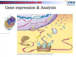
Gene expression & Analysis
Gene expression & Analysis. Where are the genes located?. Genes are located on the chromosomes. Every species has a different number of chromosomes. There are two types of chromosomes: autosomes and sex chromosomes.

Gene expression & Analysis
E N D
Presentation Transcript
Where are the genes located? • Genes are located on the chromosomes. • Every species has a different number of chromosomes. • There are two types of chromosomes: autosomes and sex chromosomes
Genes are located on the chromosomes which are found in the nucleus of a cell. • When a cell is undergoing cell reproduction, the chromosomes are visible. Chromosomes appear when the chromatin condenses and become visible. • Most of the time (90%) the genetic material in the form of chromatin. • A genome is the complete genetic information contained in an individual. • (gene + chromosome)
What is gene expression? Gene expression is the activation of a gene that results in a protein.
Gene expression takes place differently in prokaryotes and eukaryotes. • Prokaryotes • No membrane bound organelles (nucleus) • More primitive organisms • Only one circular chromosome • Bacteria are the only organisms that are prokaryotes. • Eukaryotes • Membrane bound organelles ( specialize in function –nucleus, mitochondria, chloroplast) • Chromosomes are in pairs and not circular • All organisms that are not bacteria: protist, fungi, plants and animals
DNA in eukaryotes has regions of coding and noncoding DNA. The regions of DNA that code for proteins or traits are called EXONS, while the regions that do not code for proteins are called INTRONS. cytoplasm
In Eukaryotes, following mitosis or meiosis, DNA recoils but certain regions remain relaxed for transcription. The areas of relaxed DNA are called euchromatin. • Transcription is the Reading of the DNA and Changing the code to mRNA. • Translation is changing The mRNA into a trait by Using tRNA to interpret the mRNA.
Translation • RNA • Single stranded • Does not contain thymine but has uracil instead. • tRNA carries 3 base pair code for specific amino acid. • Amino acids compose polypeptid chains. • One or more polypeptide chains compose a protein • proteins provide the “blueprints” for our characteristics and functions.
Southern hybridization 핵산분리 제한효소처리 Agarose 전기영동 Gel 전처리 Southern blot Hybridization & detection
Southern Blotting 원리 Southern(1975)에 의해 개발된 방법으로, 그 순서를 요약하면 다음과 같다. - 해석할 DNA를 적당한 제한효소로 절단한다. - 절단된 DNA를 agarose gel 전기영동에 의해 분자량의 순으로 분획한다. - 변성된 DNA를 agarose gel로부터 nitrocellulose filter에 흡착시켜 고정시킨다.- 이렇게 준비된 filter와 방사선 동위원소로 표지한 probe를 수용액 중에서 hybridization - Filter를 X선 필름에 노출시켜 방사선으로 감광된 band를 검출함으로써 목적하는 DNA 의 존재를 확인하는 것이다.
Southern Blotting 실험방법 1. DNA의 절단목적의 DNA를 적당한 제한효소로 절단한다.2. Agarose gel 전기영동Agarose gel은 두께 5 ~ 8mm 정도가 적당하며, 크기는 DNA분석용 gel보다 큰 것이 좋다. Agarose gel 전기영동은 보통 사용하는 방법과 같다.3. DNA 단편을 Agarose gel로부터 filter에 흡착DNA 단편들을 Agarose gel로부터 filter에 흡착하는 방법으로는 1) capillary transfer2) Electrophoretic transfer3) Vacuum transfer 등의 3가지가 있다. 여기에서는 가장 널리 쓰이는 capillary transfer에 대하여 설명한다. gel이 두꺼운 경우, 용액이 gel로 침루하는 시간이 다르기 때문에 두께 5 ~ 8mm 정도의 gel을 기준으로 한다. 4. Hybridization hybridization에 영향을 미치는 것은, 오노, 염농도, 시간, probe의 농도 등이다. 그러 나 특별한 경우를 제외하고는 온도는 65°C, 0.5 ~ 1.0M Nacl 농도에서 20시간 정도로 한다. Hybridization 방법은, 사용하는 probe의 종류(DNA, 합성 oligonucleotide, RNA) 따라 다르다, 그러므로 여기서는 가장 기본이 되는 DNA probe를 사용한 경우를 설 명하고자 한다.(1) 건조된 filter를 내열성 비닐포에 넣고 10mL의 prehybridization 용액을 가하여 적신 후, 기포를 제거하고 끝부분은 접착기로 봉한다.(2) 65°C, water bath에서 2 ~ 3시간 incubation한 후 비닐포의 한쪽 끝을 약간 절단하여 prehybridization 용액을 제거한다.(3) 5mL 정도의 hybridization용액 및 probe(25¡50ng)을 넣고, 기포를 제거한 후 다 시 비닐포의 한쪽 끝을 봉하여, 65 ~ 68°C에서 16 ~ 24 시간 incubation한다.(4) Hybridization이 끝난 filter를 적당한 용기에 넣어, 2X SSC-0.1% SDS로 65 ~ 68에서 10분간 천천히 흔들어 주면서 2회 세척한다.(5) 다시 1X SSC-0.1% SDS, 0.3X SSC-0.1% SDS, 0.1X SSC-0.1% SDS용액 순서 대로 65 ~ 68°C, 20분간 천천히 흔들어 주면서 세척한 후, Whatman 3 MM paper 위에 올려놓고 공기 중에서 건조시킨다.(6) Filter를 두장의 비닐랩 사이에 놓은후, 이것을 X-ray exposure cassette안에 놓 고, 접착 테이프를 이용하여 고정 시킨다.5. Autoradiography 및 X-ray flim 현상다음 (1), (3)단계는 암실, 안전등(RED FILTER)하에 실시한다.(1) 고정된 filter위에 X-ray film를 올려놓아 고정시킨후, 그 위에 intensifying screen 을 올려놓고 cassette 뚜껑을 덮는다.(2) 실온 또는 -70°C에서 2 ~ 3시간, 경우에 따라서는 1 ~ 2일 노출시킨다.(3) Film을 film holder에 걸어서 Kodak X-ray 현상액에서 2 ~ 3분 정도 담근다.(4) Film을 흐르는 물속에서 4 ~ 5분간 세척한다.(5) Kodak fixer액에 2 ~ 3분간 담근다.(6) Film을 흐르는 물속에서 10 ~ 30분 동안 세척한다.(7) Film을 걸어서 말린 후, 확인한다.
RNA 분리 1) Add 1㎖ TRIZOL 2) 5 min incubation at 15-30℃3) add 200㎕ chloroform. Shake tube vigorously by hand for 15 sec.4) 2-3 min incubation at 15-30℃5) Centrifuge at 13,200×rpm for 15 min at 2-8℃6) Transfer upper aqueous phase7) Add 500㎕ isopropyl alcohol (Precipitate the RNA) mix.8) Incubate sample at 15-30℃ for 10 min9) Centrifuge at 13,200×rpm for 10 min at 2-8℃→ pellet 없을 땐 1번 더 spin.10) Remove supernatant(빠르게 제거)11) Wash the RNA pellet once with 75% EtOH 1㎖(store 가능 : -20℃)12) Mix the sample by vortexing and Centrifuge at 13,200×rpm 5min at 2-8℃13) Dry(air dry for 5min)14) Add DEPC H2O 25㎕
Northern Blotting 원리 1975년 Southern EM이 제한효소를 절단해서 agarose gel 전기 영동으로 분획한 DNA 단편을 직접 Nitro-cellulose filter에 옮기는 방법을 개발했다. 이 Southern blot는 유전자 해석 수단으로서 아주 유용하기 때문에 그 후 널리 이용되고 있다. 1977년 Stark는 agarose gel 전기 영동으로 분획한 RNA를 filter에 옮기는 방법을 개발했다. 이 방법은 DNA에 대한Southern blot 와 대비시켜 Northern blot로 부르게 되었다. 기본적으로는 Southern blotting 과 동일하다. RNA를 전기 영동한 후 filter상에 고정하여 DNA(또는 RNA)를 probe로 하여 hybridization 해서 목적의 RNA를 검출한다. 그러나 RNA는 이차구조를 만들기 쉽기 때문에 gel 중에서 변성된 상태로 전기 영동을 시행한다. (1) 취급에 따른 일반적 주의 RNA에서 바른 정보를 얻기 위해서는 RNA를 가능한 한 분해되지 않은 상태로 조제하지 않으면 안된다. 특히 RNase에 의한 분해에 주의할 필요가 있다. RNase의 오염으로부터 시료를 지키기 위하여 고무장갑, disposable 제품, Autoclave, 건열 멸균, RNase 저해제 등을 이용한다. 유리기구, teflon제 기구는 세정후 증류수로 잘 헹구어서 건열한다. 전기 영동조 등 건열할 수 없는 기구는 세정 후 5%의 과산화수소수에 1시간 담그고 증류수로 잘 헹군다. 물은 Autoclave하여 사용한다. 시약은 autoclave 하든가 할 수 없는 것은 여과 멸균한다. 또 RNA는 DNA 와 달리 alkali에서 가수 분해되기 쉽다. 완충액 등의 pH가 alkali로 되지 않도록 주의 할 필요가 있다. (2 ) 추출조제 total RNA 나 poly(A)+RNA를 이용한다. RNA를 조제한 경우, 그 중 80% 정도가 rRNA이며 mRNA 중의 정보를 얻기 위해서는 가능한 한 rRNA의 혼입을 방지하는 것이 좋다. 목적의 mRNA가 적다고 생각되는 경우는 RNA를 Oligo(dT) column 처리를 한다. mRNA는 poly(A) 구조를 3`말단에 가지고 있기 때문에 이 조작으로 mRNA를 수 십배로 농축 정제가 가능하다. 그 외 단백질 혼입도 가능한 한피하지 않으면 안된다.
1. agarose gel의 제작(1% agarose, 1 x MOPS, 2.2mol/l formaldehyde) 2. RNA sample의 조제(RT-PCR RNA 준비과정과 동일 ) 3. 전기영동(UV 조사) - 100 - 120V, 2 - 3시간(2 - 3분간) 4. gel을 꺼내고 10 x SSC에 담근다 - 20분 - 1시간 (NaCl, sodium citrate) 5. transfer - 하룻밤 overnight 6. 건열 처리 - 80℃, 30분 - 2시간 7. prehybridization - 42℃, 2 - 3시간 8. hybridization - 42℃, 하룻밤 overnight 9. 세 정 - 1 x SSC, 0.1% SDS, 실온, 20분간 1회 0.2 x SSC, 0.1% SDS, 68℃, 20분간 3회 10. autoradiography Northern Blotting 실험방법
Detection of alternative splicing by Northern blotting • Northern blotting can be used to detect specific RNAs in complex mixtures. • Southern blotting detects specific DNA fragments. • Western blotting (immunoblotting) detects specific proteins with antibodies. RNA mixture Transfer solution RNA Question: You are using Northern blotting to analyze two mRNA samples derived from fibroblasts and hepatocytes. What will you see if you use a probe made from exon EIIIB of the fibronectin gene? What about using a probe made from the exon next to EIIIB?
RT-PCR 과정 ■ RT-PCR은 세가지 과정 (1) RNA 분리 과정 (이 과정은 Northern Blot을 하기 전에 시행해야 하는 동일한 과정이다) (2) cDNA 합성 과정(reverse transcription) (3) PCR amplification (이 과정은 Genomic DNA로부터 특정 유전자 부위를 증폭시키는 과정과 같다) 위의 세가지 과정으로 진행된다. mRNA로부터 reverse transcriptase를 이용하여 cDNA를 제조하는 방법에는 어떤 oligonucleotide를 primer로 사용하는가에 따라 세가지 방법 즉,(1) Antisense primer(3' 쪽 유전자에 특이성을 지닌 primer)를 이용하여 특정 부위 cDNA 제조(2) Random hexamer를 이용하여 전체 mRNA에 상보적인 cDNA 제조(3) Oligo dT primer를 이용하여 전체 mRNA에 상보적인 cDNA 제조가 있다. 여기서는 oligo dT primer를 이용한 방법을 사용하였다.또한 Northern Blot과 마찬가지로 cDNA가 만들어질 때까지는 RNase에 의한 오염 방지에 신경을 써야 한다.
RT-PCR 원리 RT-PCR이란 P.Seeburg(1986)에 의해 RNA를 찾고 분석하는데 도입된 방법으로 mRNA(messenger RNA)로부터 reverse transcription 과정을 통해 얻어진 cDNA(complementary DNA)를 PCR로 증폭하는 방법이다. 이러한 방법은 RNA 검사의 sensitivity를 높이고 소량의 RNA로부터 염기서열을 분석할 수 있게 하였다. 맨 먼저 reverse transcriptase로 RNA를 complementary DNA(cDNA)로 역전사합니다. 이때 쓰이는 primer는 downstream primer뿐입니다. 왜냐하면 RNA는 single strand이기 때문입니다. 그후 생성된 cDNA에서 일반적인 PCR의 과정을 밟아서 유전자를 증폭합니다.
그러면 downstream primer는 어떻게 만들까요. 크게 다음과 같은 세가지 방법이 알려져 있습니다. mRNA 뒤에 poly(A) tail이 붙는 것을 응용한 oligo(dT) primer, gene specific primer, 그리고 여기저기 붙는 random primer입니다. Oligo(dT) primer를 쓰면 분리한 RNA 중 mRNA는 모두 cDNA로 만들어집니다. Gene specific primter를 사용하면 target gene만 cDNA로 만들어지겠지요. 그리고 random primer(주로 hexamer)는 염기서열이 일정치 않지만 길이가 고정된 primer입니다. 이런 primer를 넣고 반응시키면 길이도 다양하고 모든 RNA(rRNA, tRNA 포함)가 모두 cDNA로 만들어지겠지요.
DNA synthesis is done by an enzyme (DNA polymerase) adding nucleotides to the 3’-end of a primer DNA chain
Polymerase Chain Reaction (PCR)-1 A pre-defined DNA sequence in the genome can be greatly amplified by repeated Polymerization cycles using 2 primers which hybridize to the ends of the target DNA. In each cycle, the amount of target DNA is doubled. After 10, 20 and 30 cycles, there is a 1000-, million- and billion-fold amplification respectively.
Polymerase Chain Reaction (PCR)-2 • Each PCR cycle • has 3 steps- • Melting of DNA • b. Hybridization of primer • c. DNA synthesis
Secondary antibody Western blot • Antigen- Antibody reaction을 이용하여 여러 단백질 혼합물 중에서 원하는 특정 단백질만을 찾아내는 방법 Primary antibody Block solution Nitrocellulose membrane antigen
From Gel to Blot • Polyacrylamide Gel Electrophoresis: • Break protein complexes into individual proteins • Separates protein samples based on size • Western Blot Analysis: • Transfer the proteins to a nitrocellulose membrane • More stable and permanent • Identifies proteins by immunodetection: using specific antibodies against the protein of interest
Prepare for Electrophoretic Transfer • Place the closed and locked cassette in the electrode module • Add the frozen Bio-Ice cooling unit and place in tank • Fill the tank with buffer • A stir bar can be added to help maintain the ion and temperature distribution in the tank even
Transfer Proteins from the gel to the nitrocellulose membrane30 minutes100VBlotting buffer 1x Tris glycine with 20% ethanol Electric Current
Discard blocking solution • Pour 10ml of primary antibody onto the membrane and gently rock for 10 minutes • Primary antibody will bind to the myosin light-chain • Quickly rinse membrane in 50ml of wash buffer and discard the wash buffer • Add 50ml of wash leave for 3 minutes on the rocking platform Add the Primary Antibodyanti-myosin light-chain Wash
Discard wash solution • Pour 10ml of the secondary antibody onto the membrane and gently rock for 10 minutes • Secondary antibody will bind to the primary antibody • Quickly rinse membrane in 50ml of wash buffer and discard the wash buffer • Add 50ml of wash leave for 3 minutes on the rocking platform • Western Blot animation Add Enzyme-linked Secondary AntibodyWash
Transfection and Protein localization
Exploring protein function 1) Where is it localized in the cell? Approaches: a) Make antibodies - immunofluorescence b) “Express” the protein in cells with a tag Fuse to GFP 2) What is it doing in the cell? Approaches: a) Reduce protein levels - RNA interference b) Increase protein levels “over-express” c) “Express” mutant versions Transfection!!!!
Transfection = Introduction of DNA into mammalian cells Gene is transcribed and translated into protein = “expressed”
Direct introduction of the DNA Electroporation - electric field temporarily disrupts plasma membrane Biolistics (gene gun)- fire DNA coated particles into cell Microinjection
Infection: Use recombinant viruses to deliver DNA Retroviruses Adenoviruses Virally-mediated introduction of the DNA
Carrier-mediated introduction of the DNA Positively charged carrier molecules are mixed with the DNA and added to cell culture media: Calcium Phosphate DEAE Dextran liposomes micelles Carrier-DNA complexes bind to plasma membrane and are taken up
Types of Transfection Transient: Expression assayed 24-48 hours post transfection Stable: Integration of the transfected DNA into the cell genome - selectable marker like neomycin resistance required “stably transfected” cell line
DNA “expression” vector transfected: Insert gene in here For expression in cells Polyadenylation site GFP CMV Promoter SV40 Promoter To generate stable cell line pCMV/GFP Ampicillin resistance Neomycin resistance For amplification of the plasmid in bacteria Polyadenylation site pUC Bacterial origin of replication
Three ways to make Green fluorescent protein “GFP” fusion constructs: PROTEIN X GFP GFP PROTEIN Y PROTEIN GFP Z
EXPERIMENT: Transfect unknown GFP fusion protein Protein X, Y or Z Visualize GFP protein fluorescence by fluorescence microscopy in living cells Counter-stain with known marker to compare localization patterns inliving cells = “vital stain”