Download
1 / 47
590 likes | 1.18k Vues
IDIOPATHIC NEPHROTIC SYNDROME IN CHILDREN. Dr. Hala Wannous pediatric nephrologist damascus university faculty of medicine children ’ s hospital. Introduction. Definition of NS Etiology of NS Pathology of NS Pathophysiology of NS Clinical Manifestations of NS Complications of NS

E N D
IDIOPATHIC NEPHROTIC SYNDROME IN CHILDREN Dr. HalaWannous pediatric nephrologist damascus university faculty of medicine children’s hospital
Introduction • Definition of NS • Etiology of NS • Pathology of NS • Pathophysiology of NS • Clinical Manifestations of NS • Complications of NS • Laboratory Data • Diagnosis • Treatment
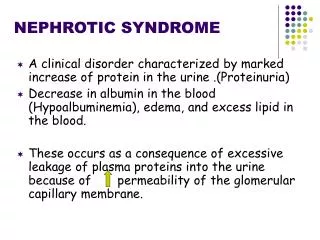
Definition Nephrotic syndrome (NS) results from increased permeability of Glomeulrar basement membrane (GBM) to plasma protein. It is clinical and laboratory syndrome characterized by massive proteinuria, which lead to hypoproteinemia ( hypo-albuminemia), hyperlipidemia and pitting edema.
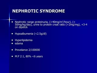
Nephrotic Criteria: *Massive proteinuria : qualitative proteinuria: persistent 3+ or 4+ urinalysis for protein, Or, urinary protein (mg)/urinary creatinine (mg) ratio in a spot sample >2 quantitative proteinuria : 24 hr urine collection for protein: ≥40 mg/m2/hr , or ≥ 50 mg/kg/24 hr *Hypoalbuminemia: serum albumin : < 2.5 g/dl. *Hyperlipidemia : serum cholesterol : > 5.7 mmol/L (>200 mg/dl) *Edema: pitting edema in different degree, often massive generalized oedema
Etiology of NS (Classification): • A- Idiopathic NS (INS): majority INS is defined by the combination of a nephrotic syndrome and non-specific histological abnormalities of the kidney. The cause is still unclear up to now. Recent 10 years, increasing evidence has suggested that INS may result from a primary disorder of T– cell function. Accounting for 90% of NS in child. mainly discussed. • B-Secondary NS: NS resulted from systemic diseases, such as anaphylactoid purpura, systemic lupus erythematosus, HBV infection. • C-Congenital NS: rare First 3 months or first year of life, only treatment renal transplantation.
Secondary NS • Primary glomerular disease : Membranous Nephropathy (MN), Membranoproliferative glomerulonephritis(MPGN), Berger disease,Cresent glomerulonephritis . • Drug,Toxic,Allegy: mercury, snake venom, vaccine, penicillamine, Heroin, gold, NSAID, captopril, probenecid, volatile hydrocarbons • Infection: HBV, HCV,HIV, shunt nephropathy, reflux nephropathy, leprosy, syphilis, Schistosomiasis, hydatid disease • Autoimmune or collagen-vascular diseases: SLE, Hashimoto’s thyroiditis, Vasculitis • Metabolic disease: Diabetes mellitus • Neoplasma: Hodgkin’s disease, carcinoma ( renal cell, lung, neuroblastoma, breast, and etc) • Genetic Disease: Alport syn, Sickle cell disease, Amyloidosis, Congenital nephropathy (nephronic reduction) • Others: Chronic transplant rejection, congenital nephrosclerosis, HUS
Idiopathic NS (INS): Pathology Minimal Change Nephrotic Syndrome (MCNS):about 85% The glomeruli appear normal basically, or show a minimal increase in mesangial cells and matrixunder light microscopy. Findings on immunofluorescence microscopy are typically negative. Under Electron microscopy : fusion of the foot processes of the podocytes Mesangial proliferative glomerulonephritis:about 5% Focal segmental glomerulosclerosis (FSGS): 7-10%
Scanning electron microscopy showing the normal aspect of podocytes with their foot processes on the left, and their effacement in minimal change disease on the right
A representation of fine structure of glomerular filter as visualized by electron microscopy. Cross-section through the center of the glomerulus.
A representation of fine structure of glomerular filter as visualized by electron microscopy. Segment of glomerular capillaries
Pathophysiology: Idiopathic nephrotic syndrome is associated with complex disturbances in the immune system, especially T cell–mediated immunity. The Main Trigger Of Idiopathic Nephrotic Syndrome and Fundamental and highly important change of pathophysiology : Proteinuria
Pathogenesis of Proteinuria: • Increased permeability of the glomerular capillary wall for proteins due to loss of negative charged glycoprotein. • On biopsy, the extensive effacement of podocyte foot processes (the hallmark of idiopathic nephrotic syndrome) suggests a pivotal role for the podocyte. • Type of proteinuria:- A-Selective proteinuria: where proteins of low molecular weight(LMW), such as albumin, are excreted more readily than protein of high molecular weight(HMW) . B-Non selective : LMW+HMW are lost in urine
Pathogenesis of hypoalbuminemia: *Due to hyperproteinuria----- Loss of plasma protein in urine mainly the albumin. *Increased catabolism of protein during acute phase.
Pathogenesis of hyperlipidemia: *Response to Hypoalbuminemia → reflex to liver --→synthesis of generalize protein ( including lipoprotein ) and lipid in the liver ,the lipoprotein (high molecular weight) not loss in urine → hyperlipidemia *Diminished catabolism of lipoprotein: loss of lipase in urine.
Pathogenesis of edema: *Reduction in plasma colloid osmotic pressure↓secondary to hypoalbuminemia Edema and hypovolemia *Intravascular volume↓antidiuretic hormone(ADH) and aldosterone(ALD) water and sodium retention Edema *Intravascular volume↓ glomerular filtration rate (GFR)↓ water and sodium retention Edema Intrarenal factors: (Nephrotic kidney retains sodium), may be involved in the formation of edema in some patients with increased intravascular volume with diminished plasma levels of renin and aldosterone.
Clinical Manifestations: In MCNS , The male preponderance of 2:1 1.Main manifestations: Edema (varying degrees) is the common symptom Local edema: edema in face , around eyes( Periorbital swelling) , in lower extremities. • Generalized edema (anasarca), ascites, pleural effusion, edema in penis and scrotum. 2-Non-specific symptoms: Fatigue and lethargy , loss of appetite, nausea and vomiting , abdominal pain , diarrhea body weight increase, urine output decrease pleural effusion (respiratory distress)
Investigations: 1-Urine analysis: a-Proteinuria: 3+ or 4 + SELECTIVE. Or, urinary protein (mg)/urinary creatinine (mg) ratio in a spot sample >2 b-24 hr urine collection for protein: ≥40 mg/m2/hr , or ≥ 50 mg/kg/24hr c- volume: oliguria (during stage of edema formation) d-Microscopically: Macroscopic hematuria is rare, occurring in 3% of patients. Microscopic hematuria is present in 20% of cases. Fat bodies, large number of hyaline casts
Investigations: 2-Blood: A-serum protein: decrease < 5.5 g/dL , Albumin levels are low (<2.5 g/dL). B-Serum cholesterol and triglycerides:high,Cholesterol >5.7 mmol/L (> 200mg/dl). C-Hemoglobin levels and hematocrit are increased in patients with plasma volume contraction. D-Serum complement: Vary with clinical type. E-Renal function : Blood urea nitrogen and creatinine concentrations are usually within the normal range, or slightly increased in relation to a modest reduction in the glomerular filtration rate (GFR).
Initial episode of Nephrotic syndrome • C3, C4, CH50, ANA, Anti DNA Antibodies, ANCA • PT, PTT, Fibrinogen, ATIII, Protein C, Protein S • Screen for tuberculosis: x-ray chest, mantoux test • Screen for urinary tract infection; urine culture and colony count. • HbsAg test, HCV Antibodies, HCV PCR, HIV Antibodies. Steroid therapy is started after the infection is cleared (for TB, after 3-4 weeks of 2 anti TB drugs).
Kidney Biopsy: Considered in: • 1- Children less than 1 year, or more than 11 years of age • 2- Macorscopic Hematuria • 3- PersistentHypertension • 4- Persistent renal failure (raised BUN, Creatinine) without hypovolemia • 5- Low serum C3 levels (Secondary NS) • 6- Steroid resistant NS • 7- Frequent relapsing NS,before considering alternative therapy, namely anticalcineurin agents.
Differential Diagnosis of NS: D.D of generalized edema:- • 1- Protein –losing enteropathy • 2- Hepatic Failure. • 3- Protein energy malnutrition • 4- Acute and chronic GN • 5- urticaria? Angio edema
Complications of NS: 1-Infections: Infections is a major complication in children with NS. It frequently trigger relapses. Nephrotic patients are liable to infection because : A- loss of immunoglobins in urine, and an impaired synthesis. B- Impaired T lymphocyte function. C- The edema fluid act as a culture medium. D- Use corticosteroids and immunosuppressive agents. E- Malnutrition The common infection : URI, peritonitis, cellulitis, and UTI may be seen. Organisms: encapsulated (Pneumococci, H.influenzae), Gram negative (e.g E.coli), and Viral infections (Chickenpox).
Complications … Vaccines in NS: polyvalent pneumococcal vaccine (if not previously immunized)at disease onset, or when the child is in remission and off daily prednisone therapy. Children with a negative varicella antibodies titer should be given varicella vaccine. Varicella vaccination is safe and effective if the child is in remission even if he is on low-dose alternate day steroids.
Complications….. 2-Hypercoagulability (Thrombosis): Hypercoagulability of the blood leading to venous or arterial thrombosis. • Hypercoagulability in Nephrotic syndrome caused by: 1- Higher concentration of I, II, V,VII, VIII, X, XIII, and fibrinogen 2- Lower level of anticoagulant substance: antithrombin III, protein C, protein S. 3- decrease fibrinolysis. 4- Higher blood viscosity (Hypovolemia) 5- Increased platelet aggregation 6- Overaggressive diuresis 7- Infection
Complications….. 3- Acute Renal Failure : pre-renal and renal. 4- cardiovascular disease : Hyperlipidemia, may be a risk factor for cardiovascular disease. 5- Hypovolemic shock 6- Others: growth retardation, malnutrition, adrenal cortical insufficiency.
Management of NS: • General (non-specific ) • *Corticosteroid therapy
General therapy: • Hospitalization: for initial work-up and evaluation of treatment. • Diet : • Protein intake of around 130–140% of the recommended daily allowance for age. • Low salt diet: only during period of edema or hypertension. • Salt-free diet: in cases of massive edema. • Restricting fluid intake: in severe edema and hyponatremia (plasma sodium concentration less than 125 meq/l). • Reduction of saturated fat is recommended. • Carbohydrates should be given preferentially as starch or dextrin-maltose, avoiding sucrose which increases lipid abnormalities.
General therapy: • Activity: usually no restriction , except massive edema, heavy hypertension, and infection. • Avoiding infection:very important. • Diuretics: • Diuretics should only be used in cases of severe edema, after hypovolemia has been corrected. • Furosemide is administered at a dose of 1–2 mg/kg. • Furosemide with Hydrochlorothiazide (HCT): 2 mg/kg/day • Spironolactone : 5–10 mg/kg • Patients with severe edema may be treated with furosemide plus albumin to increase the rate of diuretic delivery to the kidney (loop diuretics are highly protein bound). • Refractory edema with serious effusions may require drainage of ascites and/or pleural effusions.
Indication of albumininfusion therapy • Albumin : Intravenous 20% albumin infusion at a dose of 1 g/kg/dose, given over 3–4 hours. Indication: • 1- Severe edema • 2- Ascites • 3- Pleural effusion • 4- Genital edema • 5- Hypovolemia Followed immediately by furosemide (1–2 mg/kg/dose), after exclude hypovolemia.
General therapy: • Anticoagulant : • Prophylactic warfarin therapy may be given to high risk patients with : Plasma albumin concentration below 20 g/l, with: fibrinogen level over 6 g/l, or an antithrombin III level below 70% of normal. • Patients at risk may also be treated with low dose aspirin and dipyridamole. • Treatment of Hyperlipidemia: Statins are effective. • Anti Hypertension Drugs: An angiotensin converting enzyme inhibitor is preferred. • Vaccination • Preventive treatment with vitamin D and Calcium supplements.
Corticosteroid—prednisone therapy: The standard regimen : Prednisone at a dose of 60 mg/m2/day (maximum 80 mg/day), divided into 2 doses for 4 consecutive weeks . After complete absence of proteinuria (remission), prednisone dose should be tapered to 40 mg/m2 given every other day as a single morning dose for 4 weeks [ International Study of Kidney Disease in Children (ISKDC) ]. There is emerging evidence that children who receive 3 months or more steroid therapy at disease presentation appear to have a significantly higher relapse-free rate at 12 months post- presentation than those who receive the standard regimen. Thus, the alternate-day dose is slowly tapered and discontinued over the next 2-3 months.
Treatment of relapse in NS: Many children with nephrotic syndrome(50-70%) will experience one or more relapses: (proteinuria>50 mg/kg/day or Albustix +++ for 3 consecutive days after having been in remission). Treatment: Prednisone at a dose of 60 mg/m2/day (maximum 80 mg/day), divided into 2 doses until the child enters remission(proteinuria˂5 mg/kg/day or urine trace or negative for protein for 3 consecutive days). The prednisone dose is then changed to alternate- day dosing and tapered over 1-2 months.
According to response to prednisone therapy: 1- Steroid Sensitive 2- Steroid Dependent 3- Steroid Resistant Steroid Sensitive : complete remission achieved with steroid therapy (More than 95% of children with minimal change disease, and less than 20% of patients with FSGS). Steroid Dependent: ≥ 2 consecutive relapses during corticosteroid therapy, or within 14 days after cessation of therapy. Steroid resistant: failure to achieve remission following 4 weeks prednisone 60 mg/m2/day, followed by 3 pulses of methylprednisolone. Frequent relapsing: 2 or more relapses within the first 6 months after the initial remission, or 4 or more relapses within a period of 1 year.
. hyperglycemia myopathy peptic ulcer poor healing of wound. Hirsutism, striae, acne Thromboembolism Hypertension Susceptibility to infections obesity Side Effects With Long Term Use of Steroids (Steroid toxicity): • Stunted growth • Cataracts • Pseudo tumor cerebral • Psychosis • Osteoporosis • avascular necrosis of bone • Cushingoid features • Adrenal gland suppression
Major therapeutic challenges in NS are: Frequent relapses/ steroid - dependent cases First 2-3 relapses are treated with short courses of oral prednisone 60 mg/m2/day, till remission occurs followed by alternate days single dose for 4 weeks. Since repeated courses of high dose steroids cause more steroid toxicity than alternate day regimes given for 6-12 months, thus after the 3rd relapse within 6 months (frequent relapser), or steroid - dependent cases are subsequently treated with oral prednisone used in as low dose as possible on alternate days to maintain sustained remission without major side effects. Most children tolerate 0.5 mg/kg of prednisone on alternate days without side effects and maintain protein free urine.
Alternative agent: • When can be used: • Steroid-dependent patients, frequent relapsers, and steroid-resistant patients. • Cyclophosphamide Plus steroids • Cyclosporin A • Tacrolimus • Mycophenolate mofetile (MMF)
Indications for Alternative Therapy in a steroid responsive NS: • Relapse occurs on prednisone dosage >0.5 mg/kg / on alternate days plus one or more of the following :A- Unacceptable side effects of corticosteroid therapyB- High risk of toxicity: boys approaching puberty, or diabetes.C- Unusually severe relapses with hypovolemia, thrombosis, severe sepsis, or acute renal failure.D- Inadequate facilities for follow-up or concern about compliance. • Relapse on prednisone dose > 1 mg/kg on alternate days. • Drugs used for alternative therapy are introduced after inducing remission with oral prednisone therapy whilst tapering steroids. the details for dosage, duration and side – effects of these drugs is given in the following table :
THE END…. THANK YOU….