Download
1 / 65
1.3k likes | 2.24k Vues
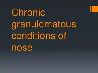
Granulomatous Disease of Nose. Granuloma consists of macrophages, epitheloid cells, and multinucleated giant cells, Blood stained discharge with crusting suggests granulomatous disease. Granulomatous conditions affecting nose and sinuses.

E N D
Granuloma consists of macrophages, epitheloid cells, and multinucleated giant cells, • Blood stained discharge with crusting suggests granulomatous disease.
Granulomatous conditions affecting nose and sinuses Infective Bacteria – Tuberculosis - Mycobacterium tuberculosis Leprosy - Mycobacterium leprae Rhinoscleroma - Klebsiellarhinoscleromatis Syphilis - Treponemapallidum Actinomycosis - Actinomycesisraeli Fungal- Rhinosporidiosis - Rhinosporidiosisseeberi Inflammatory- Wegener’s granulomatosis, Sarcoidosis, Churg-Strauss Syndrome, Cholesterol granuloma, Giant cell granuloma Neoplastic - T-cell lymphoma(midline lethal granuloma)
Tuberculosis • Poverty, overcrowding, homeless, immunocompromised. • Cutaneous form by M. tuberculosis and M. bovis. • Nasal tuberculosis caused by M. tuberculosis. • Nasal infection due to • direct inoculation by nose picking and finger nail trauma, • by open pulmonary TB, • by haematogenous dissection of primary infection elsewhere.
3 forms seen- nodular, ulcerative are seen in nose and a localised form in paranasal sinus. • Ulcerative form • usually involves cartilaginous nasal septum or anterior end of inferior turbinate, • presents with nasal obstruction, crusting, discharge and epistaxis, • unilateral or bilateral, • complication: can lead to cartilaginous septal perforation.
Nodular form also called lupus vulgaris- • Lupus typically involves normal skin and mostly in the head and neck with nose being predominant target, • begins in the vestibule and extends to adjoining skin and mucosa, • direct inoculation into dermis usually due to previous exposure. • Seen as glistening, soft, tiny, reddish-brown or skin-colored gelatinous papules or nodules (apple jelly nodules),
blanching manoeuvres, makes them prominent as surrounding area blanch while nodules remain relatively unaffected, • nodules coalesce and break down to form characteristic ulcers with a pale granular base and undermined edges or • disease may become hypertrophic with tumour-like outgrowth and deep tissue infiltration. • complication- can lead to scarring and deformity, perforation, chronic vestibulitis, extensive disruption of nasal cartilage, can also undergo malignant transformation.
Sinus granuloma- • presents with a diffuse swelling and multiple discharging sinuses in supraorbital region sec. to osteomyelitic involvement of frontal bone, • On CT and MRI, appear non-specific and demonstrate soft tissue mass with or without bone destruction, • diagnose by external or endoscopic biopsy.
Histopathology reveals typical dermal tubercles consisting of epitheloid cells, giant cells, and central caseation necrosis.
Diagnosis • Biopsy, • Microscopic examination for AFB . • Culture by Lowenstein-Jensen slope takes 6-8 weeks, culture by BACTEC system. • Manteaux test, • PCR, • Chest X-ray, • Treatment- 1st line 4 drug regime and 2 drug. 2nd line based on C/S.
Leprosy • Chronic granulomatous disease caused by M. leprae, • seen in 10-20 yrs age. • disseminated viz nasal secretions, route is URT or skin, • organism invades nerves and reside in Schwann cells. • based on immune status, clinical, histology and microbiology features, 2 forms recognised. • Tuberculoid type- has good host resistance, non-infective and localised. • Lepromatous type- has poor host resistance, infective. • systemic cases show involvement of skin, nerves, URT, eyes and testis.
Tuberculoid type- • solitary lesions causing anaesthetic cutaneous patches with involvement of sensory or motor nerves with possible paralysis of muscles. • skin of nasal vestibule can be involved but not mucosa Borderline type- no mucosal involvement.
Lepromatous type- diffuse infiltration of skin, nerves, mucosal surfaces, nasal mucosal involved early. C/F- Infection starts in anterior part of nasal septum and anterior end of inferior turbinate. Epistaxis, nasal obstruction, crust formation, blood-stained discharge with infectious bacilli. Red and swollen mucosa, shows nodular infiltration around nose, pinna, and chin. Complications- Atrophic rhinitis, septal perforation, dorsal saddling in late stages as cartilage involved, hyposmia.
Parameters for the diagnosis: • hypopigmented or reddish skin lesion with definite loss of sensations, • involvement of peripheral nerves(thickened nerves), • Slit skin smear positive for AFB. Diagnosis • Confirmed by microscopic demonstration of organism, • histopathology. • Scraping of nasal mucosa and biopsy. Treatment- rifampicin 600 mg on first of the month, clofazamine 300mg supervised on day one with clofazamine 50mg daily and dapsone 100mg daily, treatment for 12 months. In cases of deformity, corrective surgery
Rhinoscleroma • A progressive chronic granulomatous infection of nasal cavity characterised by deforming scleromatous nodules which are a consequence of inflammatory fibrosis. • Commencing in the nose and extends to nasopharynx and oropharynx, larynx, trachea, bronchi. • Common in females and middle aged. • Gram-negative bacillus Klebsiella rhinoscleromatis or Frisch bacillus, • resides intracellularly, • deficient cell mediated immunity against organism.
3 stages: atrophic stage: crust formation and foul smelling purulent discharge (mucosal atrophy), resembles atrophic rhinitis. • Granulomatous or proliferative or nodular stage- non-ulcerative nodules 1st bluish red and rubbery and later become pale and harder, fibrose and shrink around nasal vestibule, with regional lymphadenopathy, • small painless granuloma in mucosa, with subdermal infiltrates to lower part of external nose and upper lip gives a woody feel, with involvement of alar margin and nasal tip produces specific external disfigurement called ‘tapir’ nose or ‘Hebra’. • Cicatrizing stage- adhesions, stenosis, extends to nasopharynx, hard palate, trachea, bronchi
Diagnosis • biopsy with histopathologic identification of Mikulicz’s cells, foamy vacuolated histiocytes and Russell bodies, birefringent red inclusions • bacteriologically by culture of organism. • Clinically recognised in later stages, • MRI of proliferative stage shows soft tissue mass with mild to marked high signal intensity in t-1 and t-2 images. • Complement fixation test
Rhinoscleroma • Histology shows granulomatous tissue infiltrates in submucosa characterised by presence of plasma cells, lymphocytes, eosinophils, • Mikulicz cells (large foam cells with central nucleus and vacuolated cytoplasm with bacilli), • Russell bodies (resemble plasma cells with eccentric nucleus and deep eosin staining cytoplasm), • high mucopolysaccharide content in the wall of bacilli gives protection from host and antibiotics,
Treatment • streptomycin (1g/day) and tetracycline (2g/day), • or cephalosporin. Large dose for 4-6 weeks continued till 2 consecutive cultures from biopsy are proven negative. • surgical debridement and chemotherapy in granulomatous stage, • Local application 2 % acriflavine for 8 weeks, • reconstructive surgery at a later date, • steroids to reduce fibrosis.
Syphilis • Nasal involvement can occur in any age group, • contact with an infectious sexual partner, • organism usually resides and multiply in perivascular lymphatics of blood vessel wall. Leads to resultant endarteritis of small blood vessels with secondary hypertrophic changes in endothelium and luminal obliteration. • Infectious in primary and secondary forms, • Spirochaete Treponema pallidum, • IP-21days, • Congenital • Acquired - 3 stages,
Acquired • Primary lesions is at site of initial inoculation, can penetrate both normal and mucosal abrasions, • lesion is chancre and can be seen on extragenital sites such as in lips, tongue, buccal mucosa, tonsil, • in the nose, seen as firm painless nodule that may unusually be located on external nose or nasal vestibule, that breaks down to form a hard, painless ulcer with indurated margins, non tender cervical ‘rubbery’ lymphadenopathy, • secondary infection of the ulcer can lead to pain, • heals spontaneously after 2-6 weeks.
secondary syphilis seen 4-6 weeks after primary lesion, • this stage lasts few weeks • lesions seen as snail track ulcers (ulcerated lesion covered with a greyish white membrane, scraping shows pink base with no bleeding), usually seen in oral cavity, these are rarely recognised in the nose, as it hardly occurs on thin, attenuated mucous membrane, • associated with mucopurulent rhinitis, nasal crusting and vestibular fissuring.
diagnosed by appearance of other secondary lesions, development of mucous patches in pharynx, roseolar or papular mucocutaneous rash, pyrexia, shotty enlargement of lymph nodes, headache, malaise, sore throat, • lesions in mouth and pharynx are infectious.
Tertiary stage - most commonly seen in the nose, • seen 5-25years after initial infection, • lesions can be local or general, • in URT is gumma, seen on hard palate, nasal septum, tonsil, PPW larynx. • Gumma begins as subcutaneous painless nodule which breaks down to form punched out ulcer that attacks mucosa, periosteum, and bone of nose with atrophy and scarring. • External nose is initially spared though tenderness and swelling may been seen over nasal bridge.
Early symptoms include pain (worse at night), swelling and obstruction. Swelling may be diffuse or localised and offensive discharge, bleeding, crusting, anosmia. • Complication - Destruction of nasal bone and skin leads to collapse and disfigurement. Bony and cartilage portion of septum is commonly involved. perforation of affected nasal wall, saddle nose deformity. Scarring and secondary atrophic rhinitis, vestibular stenosis, perforation of hard palate,
Congenital infection occurs in 2 forms: • early and late- • Early- In infant, snuffles seen, begins around 3rd week to 3 months after birth, at first appears as simple catarrhal rhinitis. Becomes purulent with secondary fissuring and excoriation of nasal vestibule and upper lip, interferes with suckling and nutrition. • family history of syphilis, miscarriage, still birth, other stigmata like Hutchinsons’ incisors, Moon’s molars, interstitial keratitis, corneal opacities, SN deafness.
Late manifests around puberty, similar to tertiary acquired stage, • Gummatous and destructive lesions occur most commonly, mucous membrane, periosteum and bone affected. Resulting ulceration and destruction lead to features of secondary atrophic rhinitis and sinking of nasal bridge, produce saddle nose deformity,
Diagnosis: In primary, smears from ulcer examined by dark-ground illumination or after staining show the spirochaete, -serology includes VDRL(venereal disease reference laboratory), TPHA(t.pallidum haemagglutination test), FTA-ABS(fluorescent treponemal antibody test). -biopsy. Secondary stage confirmed by serology -dark field examination of lymph node aspirates or tissue specimens by silver stains or immunofluorescence. -VDRL. Tertiary stage by biopsy, -serology by VDRL, FTA, TPHA, T.pallidum immobilisation test
Treatment: • In primary and secondary stage, penicillin in single or divided dose as benzathine penicillin- 2.4 mega units, • in tertiary stage, 7.2 mega units in divided dose of 2.4 mega units at 7 to 14 day intervals. • Local toilet to clear crusts and regular cleaning of nasal passage by alkaline nasal douches 1-3 times a day, • debridement and topical yellow mercuric oxide ointment to nasal vestibule. • Surgery for cosmesis in inactive stage • In snuffles, restore airway for suckling by remove nasal discharge by gentle suction and irrigation and use of normal saline drops, hyperextend the head before feeding,
Rhinosporidiosis • Chronic granulomatous infection affecting nasal mucosa, ocular conjunctiva and other mucosa like nasopharynx, lip, palate, conjunctiva, epiglottis, larynx, trachea, bronchi, and skin. • Caused by Rhinosporidium seeberi. • A fungal infection characterised by polypoid or nodular lesions which at times involve nasal surface. In tissues, form abundant large thick walled sporangium structure containing large number of endospores. • Seen in exposure to stagnant pools of contaminated fresh water, • Males, 15-40 years
Causes production of large sessile or pedunculated lesions that affect one or both nostrils, • Seen as papular or nodular smooth-surfaced lesions that become pedunculated and acquire papillomatous or proliferative appearance, lesions are pink, red or purple in color, continue to enlarge in size. Very vascular and bleeds on touch. Surface is studded with white dots representing sporangia of fungus. • Attached to septum or lateral wall. Can extend into nasopharynx and into oropharynx. • h/o blood tinged nasal discharge, nasal obstruction, epistaxis • Diagnose by histopath. • Treatment is surgical excision of lesion with or without cauterisation of base
Rhinosporidiosis A section of human nasal polyp was stained with Periodic acid-Schiff (PAS). The thick walls of immature R. seeberi trophocytesstain with PAS (pink), and the spherical organisms are surrounded by inflammatory cells, largest sporangia are filled with spores
Rhinosporidiosis • Clinical photograph of the patient showing nasal and oropharyngeal Rhinosporidiosis
Sarcoidosis • Chronic multi-systemic disorder of unknown aetiology which can affect any part of the body. • Seen in 3rd to 5th decade, more in females. • Causes pulmonary symptoms, Cervical lymphadenopathy which is bilateral and non-tender, skin, eyes, liver, spleen, small bones of hands and feet.
Nasal symptoms: nasal stuffiness and obstruction, crusting, blood-stained or mucopurulent discharge, facial pain, anosmia. • mucosa has characteristic granular appearance sometimes called strawberry skin. it has tiny pale submucosal granulomas against erythematous mucosa, very friable. • Septum and turbinates affected. Anterior nasal septum may perforate. Soft tissue mass or expansion of nasal bridge associated with thickening and discoloration of overlying skin maybe seen as in classic lupus pernio. • Paranasal sinus may also be affected or become secondly infected. Lymphoid hyperplasia and adenoidal enlargement- lead to OM with effusion and sleep disturbance
Diagnosis • biopsy showing noncaseating granulomas, • Haematology shows mild anaemia, leucopenia, thrombocytopenia eosinophilia, • Kveim test • Plain x-ray nasal bone show rarefaction or punctate osteolysis, • Angiotensin converting enzyme is elevated during the disease. • ESR, serum globulin, serum and urinary calcium are elevated, • Inv of LRT like Chest x-ray, CT , perfusion studies, bronchoalveolar lavage, gallium-67 scanning. X-ray shows diffuse pulmonary infiltrate with hilar adenopathy
Sarcoidosis Histological section of sarcoid tissue showing granuloma with Langhans’ multi-nucleated giant cells, epithelioid cells, lymphocytes and fibroblasts but devoid of caseation. Crystalline or calcified inclusion bodies usually seen eg. Schaumann bodies.
CT scan nasal sinuses showing blockage of the osteo-meatal complexes and mucosal thickening over the maxillary and ethmoidal sinuses
Treatment- steroids, methotrexate, hydroxychloroquine. • Local intranasal steroids, glucose and glycerine drops, nasal douching and irrigation, • systemic medication. • During active disease, cosmetic surgery is contraindicated
Wegener’s granulomatosis • Condition characterised by granulomatous inflammation involving respiratory tract and necrotizing vasculitis affecting small to medium size vessels with necrotising glomerulonephritis. • Coexistence of vasculitis and granulomas and involves airway, lung, and renal disease. • 15 to 73 years age. • Inflammatory in nature, hypersensitivity reaction with an immune response to an unknown stimulus, cANCA positive
C/F- Nasal obstruction, epistaxis, bloody crusts in nose, nasal discharge, facial pain, progressive malaise, pyrexia, weight loss. Intranasal destruction of bone and cartilage lead to perforation and nasal collapse, destruction of septum.