Download
1 / 75
780 likes | 876 Vues
Granulomas are nodular collections of macrophages and lymphocytes found in specific pathologic conditions. Recognizing this pattern is crucial as it could point to serious illnesses such as tuberculosis, leprosy, and more. Explore the causes, mechanisms, and types of granulomas in this comprehensive guide.

E N D
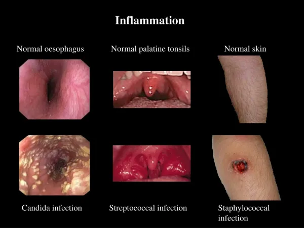
Granulomatousinflammation • A granuloma is a microscopic aggregation of macrophages that are transformed into epithelium-like cells surrounded by a collar of mononuclear leukocytes, principally lymphocytes and occasionally plasma cells.
Granulomatous Inflammation • Granuloma = Nodular collection of epithelioid macrophages surrounded by a rim of lymphocytes • Epitheloid macrophages: squamous cell-like appearance
Why is it important? • Granulomas are encountered in certain specific pathologic states; consequently, recognition of the granulomatous pattern is important because of the limited number of conditions (some life-threatening) that cause it
Granulomatousinflammation • Epithelioid cells fuse to form giant cells containing 20 or more nuclei. • The nuclei arranged either peripherally (Langhans-type giant cell) or • haphazardly (foreign body-type giant cell). • These giant cells can be found either at the periphery or the center of the granuloma.
Caseous Necrosis Epithelioid Macrophage Langhans Giant Cell Lymphocytic Rim
Granulomatous Inflammation Causes Immune granuloma: Non-immune granuloma Foreign body Splinter Suture Graft material • Bacteria • Tuberculosis • Leprosy • Actinomycosis • Cat-scratch disease • Parasites • Schistosomiasis • -Leishmaniasis • Fungi • Histoplasmosis • Blastomycosis • Metal/Dust • Berylliosis • Silicosis unknown Sarcoidosis
Granulomatousinflammation • Foreign body Granulomas: • endogenous ( keratin, necrotic bone or adipose tissue, uric acid crystals) • Exogenous (wood, silica, asbestos, silicone,suture…) • Specific chemicals: • Beryllium
Granulomatous Inflammationmechanism • What is the initiating event in granuloma formation? • deposition of a indigestible antigenic material IFN-γreleased by the CD4+ T cells of the TH1 subset is crucial in activating macrophages.
Granuloma: bacilli are inhaled by droplets Bacteria are phagocytosed by alveolar macrophages After amassing substances that they cannot digest, macrophages lose their motility, accumulate at the site of injury and transform themselves into nodular collections; the Granuloma A localized inflammatory response recruits more mononuclear cells The granuloma consists of a kernel of infected macrophages surrounded by foamy macrophages and a ring of lymphocytes and a fibrous cuff (containment phase) Containment usually fails when the immune status of the patient changes; the granuloma caseates, ruptures and spills into the airway
Langhans Giant Cell Lymphocytic Rim Epithelioid Macrophage Caseous Necrosis Granuloma
EtiologyMycobacterum tuberculosis • Mycobacteria – ‘fungus like.. • slender rods • acid fast bacilli [AFB] (i.e., they have a high content of complex lipids that readily bind the Ziehl-Neelsen [carbol fuchsin] stain and subsequently resist decolorization). • Mycobacterium bovis …..intestinal TB , milk injection • Other types • M. leprae (Hansen bacillus) ………………………..Leprosy • M. kansasii, M. avium, M. intracellulare …………..Atypical mycobacterial infections • M. ulcerans ………………………………………….Buruli ulcer
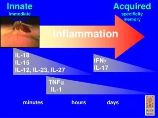
Pathogenesis of TB: Infection - Immunity
If the bacilli enter the body…… The bacilli have 4 potential fates: • (1) They may be killed by the immune system, • (2) they may multiply and cause primary TB, • (3) they may become dormant and remain asymptomatic, • (4) they may proliferate after a latency period (reactivation disease). Reactivation TB may occur following either (2) or (3) above. • (5 ) if immunosuppressed ---- Primary Progressive TB Miliary TB
TB • Primary tuberculosis [initial infection] • secondary tuberculosis [ re-activation or re-infection ]
Primary tuberculosis • Non immunized individual [initial infection] – children • Subpleural zone of lung – can be at other sites • Brief acute inflammation – neutrophils. • 5-6 days invoke granuloma formation. • 2 to 8 weeks – healing – Ghon focus (+ lymph node Ghon complex) • Develop immunity – Mantoux positive ( tuberculin test , PPD )
Primary or Ghon’s ComplexCharacteristics • initial infection • non immunized individual • 5-6 days …granuloma • 2 to 8 weeks – healing • subpleural zone…. Ghon focus • + lymph node Ghon complex • Develop immunity – Mantoux positive [ PPD ]
Secondary Tuberculosis: • Post Primary in immunized individuals. • Reactivation or Reinfection • Cavitary Granulomatous response. • Apical lobes or upper part of lower lobes – O2 • Caseation, cavity - soft granuloma • Pulmonary or extra-pulmonary • Local or systemic spread / Miliary • Vein – via left ventricle to whole body • Artery – miliary spread within the lung
Secondary Tuberculosis: • Cough, sputum, Low grade fever, night sweats, fatigue and weight loss. • Hemoptysis or pleuritic pain = severe disease
Miliary TB • Millet like – grain. • Low immunity • blood or bronchial spread • Pulmonary or Systemic types.
Diagnosis of TB • Clinical features • Depend on organ involved. • Pulmonary tuberculosis (TB): • productive cough, fever, and weight loss, night sweats.
Investigations • Patients suspected of having tuberculosis (TB) • Tuberculin skin testing (Mantoux test, PPD) • Intradermal injection of purified protein derivative ( PPD). • The response is measured as the amount of induration at 48-72 hours. • The size of induration, rather than erythema, is diagnostic. • BCG gives + result • Sputum, bronchial wash or biopsy • Acid fast smear ( ZN stain ) • cultures require weeks for growth and identification • Newer technologies, including ribosomal RNA probes or DNA polymerase chain reaction, allow identification within 24 hours. • Chest radiographs • patchy or nodular infiltrate. • may be found in any part of the lung, but upper-lobe involvement is most common
What will be your action after diagnosis? • Patients with TB should remain in isolation until sputum becomes negative;
1° TB usually involves the middle or lower lung zones and is associated with hilar adenopathy (Gohn complex). • 2 ° TB represents reactivation and typically involves the upper lungs and cavitation. • regimen RIPE—Rifampin, Isoniazid (INH), Pyrazinamide, and Ethambutol daily for eight weeks, followed by INH and rifampin for an additional 16 weeks. Give vitamin B6 to prevent INH-associated neuropathy.
Leprosy • Leprosy is a chronic infection caused by the acid-fast, rod-shaped bacillus Mycobacterium leprae. • skin • peripheral nerves
Leprosy Symptoms • skin • Painless skin patch • peripheral nerves • Loss of sensation • Wasting and muscle weakness • Foot drop or clawed hands • Ulcerations on hands or feet
Aetiology • Mycobacterium leprae • Acid fast gram-positive bacillus • cannot be cultured • The mode of transmission is unknown, probably inhalation of bacilli • incubation period is several years. • The classical method for demonstrating leprosy bacilli in lesions is a modified Ziehl-Neelsen stain. The Fite methods are the most commonly used