Inflammation
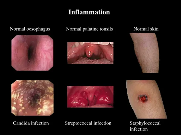
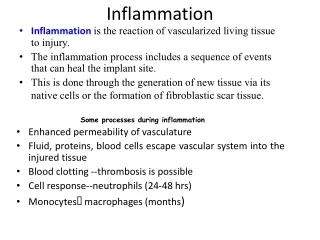
Inflammation. Acute inflammation. The cardinal signs of inflammation are rubor ( redness ), calor ( heat ), tumor ( swelling ), dolor ( pain ), and loss of function . Seen here is skin with erythema, compared to the more normal skin at the far right. .


Inflammation
E N D
Presentation Transcript
The cardinal signs of inflammation are rubor (redness), calor (heat), tumor (swelling), dolor (pain), and loss of function. Seen here is skin with erythema, compared to the more normal skin at the far right.
Peripheal blood smear. Perhaps the simplest indicator of acute inflammation is an increase in the white blood cell count in the peripheal blood, here marked by an increase in segmented neutrophils (PMN's).
Seen here is vasodilation with exudation that has led to an outpouring of fluid with fibrin into the alveolar spaces, along with PMN's.
Here PMN's that are marginated along the dilated venule wall (arrow) are squeezing through the basement membrane (the process of diapedesis) and spilling out into extravascular space.
Here is an example of the fibrin mesh in fluidwith PMN's that has formed in the area of acute inflammation. It is this fluid collection that produces the "tumor" or swelling aspect of acute inflammation.
The vasculitis shown here demonstrates the destruction that can accompany the acute inflammatory process and the interplay with the coagulation mechanism. The arterial wall is undergoing necrosis
At higher magnification, vasculitis with arterial wall necrosis is seen. Note the fragmented remains of neutrophilic nuclei (karyorrhexis).Acute inflammation is a non-selective process that can lead to tissue destruction.
A variety of inflammatory cell types may be present in inflammatory reactions, though one may predominate. Seen here are mainly neutrophils, but there are also plasma cells, lymphocytes, and macrophages. Macrophages can phagocytoze other cells as well as cellular debris. One macrophage here has "pigged out" by consuming a neutrophil, a red blood cell, and a nuclear fragment.
Here is simple edema, or fluid collection within tissues. This is "pitting" edema because, on physical examination, you can press your finger into the skin and soft tissue and leave a depression.
This example of a fluid collection, a blister of the skin, is an almost example of serous effusion.
Here is an example of fluid collection into a body cavity, or an effusion. This is a right pleural effusion (in a baby). Note the clear, pale yellow appearance of the fluid. This is a serous effusion.
The large amount of fibrin in an exudate can form a fibrinous exudate on body cavity surfaces. Here, the pericardial cavity has been opened to reveal a fibrinous pericarditis with strands of stringy pale fibrin between visceral and parietal pericardium.
Microscopically, the fibrinous exudate is seen to consist of pink strands of fibrin jutting from the pericardial surface at the upper left. Below this, there are a few scattered inflammatory cells.
Here is a purulent exudate in which the exuded fluid also contains a large number of acute inflammatory cells. Thus, the yellowish fluid in this opened pericardial cavity is a purulent exudate.
A purulent exudate is seen beneath the meninges in the brain of patient with acute meningitis. The exudate obscures the sulci.
The PMN's seen here are in alveoli, indicative of an acute bronchopneumonia of the lung. The PMN's form an exudate in the alveoli.
Numerous neutrophils fill the alveoli in this case of acute bronchopneumonia in a patient with a high fever.
Extensive acute inflammation may lead to abscess formation, as seen here with rounded abscesses (the purulent material has drained out after sectioning to leave a cavity) in upper lobe.
The white arrows mark areas of abscess formation in the upper lobe of this lung. The liquefactive necrosis of an abscess is apparent, because the purulent contents are draining out to leave a cavity.
Small abscesses are seen here. These could be termed "microabscesses" due to their small size. Abscesses can come in a variety of sizes. Perhaps the most common abscess is the pimple on the face of a teenager.
An abscess is a localized collection of PMN's. Here is a microabscess in the myocardium. The irregular dark purple center is a collection of bacteria that are the cause for this abscess.
This abscessing bronchopneumonia has numerous areas of raised, lighter tan appearance.
Microscopically, the extensive neutrophilic exudate of an acute abscessing pneumonia is seen here. Normal tissues are destroyed in the region of the abscess.
One of the morphologic patterns of acute inflammation is ulceration. This occurs on epithelial surfaces. Here the gastric mucosa has been lost, or ulcerated. A larger ulcer and several adjacent smaller ones with surrounding erythema appear at the left of center.
An esophageal acute ulcer is shown here in which the squamous mucosa has been lost. In the ulcer base are inflammatory cells and fibrin.
This patient had diabetes mellitus for many years. A transmetatarsal amputation has already been performed in this patient because of the severity of peripheral vascular disease.
Seen here is chronic endometritis with lymphocytes as well as plasma cells in the endometrial stroma. In general, the inflammatory infiltrate of chronic inflammation consists mainly of mononuclear cells (lymphocytes, plasma cells, and macrophages).
Here is chronic cervicitis. In this case the inflammation is severe enough to produce mucosal damage with hemorrhage.
Seen here in the synovium from the joint of a patient with rheumatoid arthritis are collections of dark blue lymphocytes.
Here, chronic inflammation of the bronchi has led to dilation and scarring with increased tan to white collagenous tissue.
Healing of inflammation often involves ingrowth of capillaries and fibroblasts. This forms granulation tissue. Here, an acute myocardial infarction is seen healing. There are numerous capillaries, and collagen is being laid down to form a scar. Non-infarcted myocardium is present at the far left.
At high magnification, granulation tissue has capillaries, fibroblasts, and a variable amount of inflammatory cells (mostly mononuclear
Grossly, a granuloma tends to be a focal lesion. Seen here in a hilar lymph node is a granuloma. Granulomas due to infectious agents such as mycobacteria are often described as "caseating" when they have prominent caseous necrosis.
Here are two pulmonary granulomas. Granulomatous inflammation typically consists of mixtures of cells including epithelioid macrophages, giant cells, lymphocytes, plasma cells, and fibroblasts. There may even be some neutrophils.
Langhans type giant cells are a "committee" of epithelioid macrophages. Seen here are two Langhans type giant cells in which the nuclei are lined up around the periphery of the cell. Additional pink epithelioid macrophages compose most of the rest of the granuloma.
These are epithelioid cells around the center of a granuloma. They get their name from the fact that they have lots of pink cytoplasm similar to squamous epithelial cells. Their nuclei tend to be long and stringy.