Inflammation
Inflammation. Jan Laco, MD, PhD. Inflammation. complex protective reaction caused by various endo- and exogenous stimuli injurious agents are destroyed, diluted or walled-off without inflammation and mechanism of healing could organism not survive can be potentially harmfull. Terminology.

Inflammation
E N D
Presentation Transcript
Inflammation Jan Laco, MD, PhD
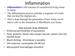
Inflammation • complex protective reaction • caused by various endo- and exogenous stimuli • injurious agents are destroyed, diluted or walled-off • without inflammation and mechanism of healing could organism not survive • can be potentially harmfull
Terminology • Greek root + -itis • metritis, not uteritis • kolpitis, not vaginitis • nephritis, not renitis • glossitis, not linguitis • cheilitis, not labiitis
Mechanisms • A) local - mild injury • B) systemic – severe injury • 3 major changes • 1. alteration – tissue change • 2. exudation - inflammatory exudate • liquid + proteins (exudate) • cellular (infiltrate) • 3. proliferation • formation of granulation and fibrous tissue • usually - all 3 components - not the same intensity
Classification • several points of view • according to length • acute × chronic (+ subacute, hyperacute) • according to predominant component • 1. alterative • 2. exudative • 3. proliferative
Classification • according to histological features • non-specific (not possible to trace etiology) - vast majority • specific / granulomatous (e.g. TBC) • according to causative agent • aseptic (sterile) - chemical substances, congelation, radiation - inflammation has a reparative character • septic (caused by living organisms) - inflammation has a protective character
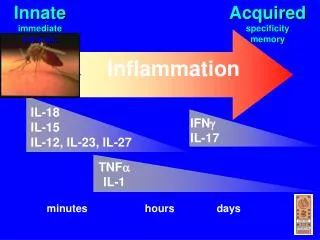
Acute inflammation • early response • important role in inflammation has microcirculation! • supply of white blood cells, interleukins, fibrin, etc.
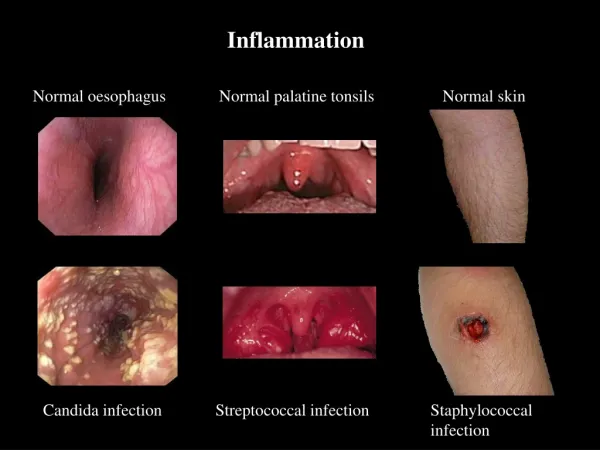
Local symptomatology • classical 5 symptoms (Celsus, 1st c. BC) • 1. calor – heat, warmth • 2. rubor – redness, erythema • 3. tumor – swelling, edema • 4. dolor - pain • 5. functio laesa – function loss/impairment
Systemic symptoms • fever (irritation of thermoregulatory centre) • TNF, IL-1 • IL-6 – high RBCs sedimentation rate (via fibrinogen) • leukocytosis - increased WBCs number • bacteria – neutrophils • parasites – eosinophils • viruses - lymphocytosis • leukopenia - decreased WBCs number • viral infections, salmonella infections, rickettsioses • immunologic reactions – “acute phase reactants“ • C-reactive protein, complement, SAA, fibrinogen, ...
Vascular changes • 1. arteriolar vasodilation (redness + warmth) • 2. increased permeability of vessels • widened intercellular junctions • retraction of endothelial cells (histamin, VEGF, bradykinin) • protein-poor transudate (edema) • protein-rich exudate • 3. endothelial injury – direct x leukocyte-dependent • proteolysis – protein leakage • platelets adhesion thrombosis
Cellular events • leukocytes margination rolling adhesion transmigration by diapedesis (in venules) • transmigration • neutrophils (1-2 days) • monocytes (2-3 days) • chemotaxis (along chemical gradient) • endogenous signaling molecules – ILs, LTs, C5a • exogenous – toxins, bacterial proteins, ... • phagocytosis (see below) • passive migration of RBCs • no active role in inflammation - hemorrhagic inflammation
Phagocytosis • 1. recognition and attachment • facilitated by opsonins (IgG, C3b) • 2. engulfment • pseudopods formation phagocytic vacuole + lysosome phagolysosome • 3. killing and degradation • oxidative burst – reactive oxygen metabolits – superoxide ion, hydrogen peroxide, hypochlorous radicals • lysosomal acid hydrolases • in highly virulent microorganisms can die leukocyte and not the microbe • in highly resistant microorganisms - persistence within macrophage - activation after many years (TBC)
Outcomes of acute inflammation • 1. resolution - restoration to normal, in limited injury • chemical substances neutralization • normalization of vascular permeability • apoptosis of inflammatory cells • increased lymphatic drainage • 2. healing by granulation tissue / fibrous scar • tissue destruction • fibrinous inflammation adhesions, fibrosis • purulent inflammation abscess formation (pus, pyogenic membrane, resorption - pseudoxanthoma cells - weeks to months) • 3. progression into chronic inflammation
Chronic inflammation • reasons: • persisting infection or prolonged exposure to irritants (intracell. surviving of agents - TBC) • repeated acute inflammations (otitis, rhinitis) • primary chronic inflammation - low virulence, sterile inflammations (silicosis) • autoimmune reactions (rheumatoid arthritis, glomerulonephritides, multiple sclerosis)
Chronic inflammation • chronic inflammatory cells ("round cell" infiltrate) • lymphocytes (T and B), plasma cells • eosinophils – parasites, allergies • monocytes / macrophages activation by various mediators - fight against invaders • B lymphocytes plasma cells, Ig production • NK cells • monocytes-macrophages specialized cells (siderophages, gitter cells, mucophages)
Morphologic patterns of inflammation • 1. alterative • poliomyelitis anterior acuta, diphtherial myocarditis • 2. exudative • 2a. serous • 2b. fibrinous • 2c. suppurative • 2d. necrotizing, gangrenous • 2e. non-purulent • 3. proliferative • primary (rare) x secondary (cholecystitis)
Morphologic patterns of inflammation • 2a. serous • excessive accumulation of fluid, few proteins • e.g. skin blister, serous membranes - initial phases of inflammation, effusions • modification - catarrhal - accumulation of mucus on mucosas - larynx • 2b. fibrinous • higher vascular permeability - exudation of fibrinogen -> fibrin • formation of pseudomembranes - fibrin, necrotic mucosa, etiologic agens, leukocytes • e.g. diphtheria - Corynebacterium, dysentery – Shigella spp., Cl. difficile • e.g. pericarditis (cor villosum, cor hirsutum - "hairy" heart) • e.g. lobar pneumonia – Str. pneumoniae • fibrinolysis resolution • organization fibrosis scar, adhesions
2c. suppurative (purulent) - accumulation of neutrophillic leukocytes - formation of pus • pyogenic bacteria - Staphylococci • interstitial • phlegmone – diffuse • abscess - localized collection • acute – border – surrounding tissue • chronic – border - pyogenic membrane • pseudoabscess – pus in lumen of hollow organ (epithelium) • formation of suppurative fistule • accumulation of pus in preformed cavities - empyema (gallbladder, thoracic cavity)
complications of suppurative inflammation • bacteremia • no clinical symptoms! • formation of secondary foci of inflamm. (endocarditis, meningitis) • sepsis = massive bacteremia • septic fever, activation of spleen, septic shock • thrombophlebitis • secondary inflammation of vein wall followed by thrombosis - embolization • pyemia - hematogenous abscesses (infected infarctions) • lymphangiitis, lymphadenitis
2d. necrotizing • inflammatory necrosis of the surface - ulcer (skin, stomach) • gangrenous - secondary modification by bacteria - apendicitis, cholecystitis - risk of perforation – peritonitis • 2e. non-purulent • round cell inflammatory infiltrate
Granulomatous inflammation • distinctive chronic inflammation type • cell mediated immune reaction (delayed) • aggregates of activated macrophages epithelioid cell multinucleated giant cells (of Langhans type x of foreign body type) • lymphocytic rim • NO agent elimination but walling off • intracellulary agents (TBC) x inert foreign bodies
Granulomatous inflammation • 1. Bacteria • TBC • leprosy • syphilis (3rd stage - gumma) • 2. Parasites + Fungi • 3. Inorganic metals or dust • silicosis • berylliosis • 4. Foreign body • suture (Schloffer “tumor“), breast prosthesis, vascular graft • 5. Unknown • – sarcoidosis, Wegener´s granulomatosis, Crohn disease
Tuberculosis – general pathology • 1. TBC nodule – proliferative • Gross: grayish, firm, 1-2 mm (milium) central soft yellow necrosis (cheese-like – caseous) calcification • Mi: central caseous necrosis (amorphous homogenous + karyorrhectic powder) + macrophages epithelioid cells multinucleated giant cells of Langhans type + lymphocytic rim • 2. TBC exudate – sero-fibrinous exudate (macrophages)
Leprosy • M. leprae, Asia, Africa • in dermal macrophages and Schwann cells • air droplets + long contact • rhinitis, eyelid destruction, facies leontina • 1. lepromatous – contagious • skin lesion – foamy macrophages (Virchow cells) + viscera • 2. tuberculoid – sterile • in peripheral nerves – tuberculoid granulomas - anesthesia • death – secondary infections + amyloidosis
Syphilis • Treponema pallidum (spirochete) • STD + transplacental fetus infection • acquired (3 stages) x congenital • basic microscopic appearance: • 1. proliferative endarteritis (endothelial hypertrophy intimal fibrosis local ischemia) + inflammation (plasma cells) • 2. gumma – central coagulative necrosis + specific granulation tissue + fibrous tissue
Syphilis • 1. primary syphilis - contagious • chancre (ulcus durum, hard chancre) • M: penis x F: vagina, cervix • painless, firm ulceration + regional painless lymphadenopathy • spontaneous resolve (weeks) scar
Syphilis • 2. secondary syphilis - contagious • after 2 months • generalized lymphadenopathy + various mucocutaneous lesions • condylomata lata - anogenital region, inner thighs, oral cavity
Syphilis • 3. tertiary syphilis • after long time (5 years) • 1) cardiovascular - syphilitic aortitis (proximal a.) • endarteritis of vasa vasorum scaring of media dilation aneurysm (thoracic aorta) • 2) neurosyphilis – tabes dorsalis + general paresis • degeneration of posterior columns of spinal cord sensory + gait abnormality • cortical atrophy psychic deterioration • 3) gumma – ulcerative lesions of bone, skin, mucosa – oral cavity
Congenital syphilis • 1) abortus • hepatomegaly + pancreatitis + pneumonia alba • 2) infantile syphilis • chronic rhinitis (snuffles) + mucocutaneous lesions • 3) late (tardive, congenital) syphilis • > 2 years duration • Hutchinson triad – notched central incisors + keratitis (blindness) + deafness (injury of n. VIII) • mulberry molars + saddle nose