Inflammation
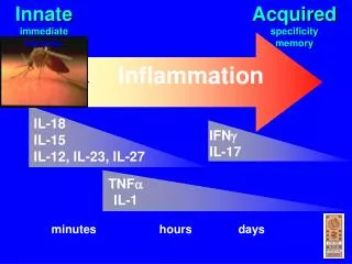
Inflammation. 沈 弘 德 台北榮總教研部. 本章大綱 Leukocyte migration Cell-adhesion molecules Mediators of inflammation The inflammatory process Anti-inflammatory agents . Lymphocyte recirculation routes. * Extravasation * Cell-adhesion molecules (CAMs) (endothelial cells & leukocytes)

Inflammation
E N D
Presentation Transcript
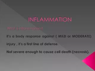
Inflammation 沈 弘 德 台北榮總教研部
本章大綱 • Leukocyte migration • Cell-adhesion molecules • Mediators of inflammation • The inflammatory process • Anti-inflammatory agents
* Extravasation * Cell-adhesion molecules (CAMs) (endothelial cells & leukocytes) - selectins - mucins - integrins - ICAMS * Receptors bind to CAMs
General structures of the four families of cell-adhesion molecules * Glycosylated proteins * Heterodimeric proteins * Expressed by leukocytes * Facilitate adherence (LAD disease) * Contain Ig/mucin-like domains * Expressed on endothelial cells * Bind to integrin/selectin molecules * Interact with sialylated carbohydrate moieties of mucin-like molecules. * Responsible for the initial stickiness of leukocytes to vascular endothelium.
Steps in neutrophil extravasation Inflammatory response ↓ cytokines, inflammatory mediators Vascular endothelium activated Increased expression of CAMs Receptor activation- induced conforma-tional change in the integrin molecules → increased affinity & adhesion Neutrophil activation by chemoattractant stimulus: chemokines, PAF, C5a, C3a, C5b67, bacterial peptides The neutrophil tumbles end-over-end along the endothelium. The neutrophil migrates through the vessel wall into the tissues.
Cell-adhesion molecules and chemokines involved in the first three steps of neutrophil extravasation adhesion rolling activation
Schematic cross-sectional diagram of a lymph node postcapillary venule with high endothelium (HEV) As many as 1.4 x 10⁴ lymphocytes extravasate every second through HEVs into a single lymph node. Each of the secondary lymphoid organs, with the exception of the spleen, contains HEVs.
Lymphocytes attached to the surface of a high-endothelial venule
Naïve T cell tend to home to secondary lymphoid tissues through their HEV regions Activation of a naïve cell occurs within secondary lymphoid tissue. (Homing receptor) (Mucin-like CAM)
Steps in extravasation of a naïve T cell through a high endothelial venule into a lymph node • Activation of • integrin molecules (a homing receptor) • G-protein-coupled • receptors • Lymphocyte-specific • chemoattractants
Mediators of inflammation • Chemokines and other mediators released by tissue • mast cells, blood platelets, leukocytes and lymphocytes. • Mediator-producing systems in plasma: • - the kinin system • - the clotting system • - the fibrinolytic system • - the complement system • Some lipids act as inflammatory mediators • Some cytokines are important inflammatory mediators
Chemokines - key mediators of inflammation
Chemokines • A superfamily of small polypeptides (90-130 aa residues) • Major regulators of leukocyte traffic • (adhesion, chemotaxis, activation) • Involved in inflammation, homeostatic/developmental • processes, angiogenesis, wound healing • Induced in response to infection • → Assembly of leukocytes at sites of infection • > 50 chemokines, possess 4 conserved cysteine residues • (C-C subgroup, C-X-C subgroup) • > 15 chemokine receptors (CC receptors, CXC receptors) • The interaction between chemokines and their receptors • is of high affinity (Ka > 109) and high specificity.
Chemokines signal through receptors coupled with heterotrimeric large G proteins • Changes in shape of leukocytes • Activation of integrins • Generation of oxygen radicals • Release of granular contents and proteases
Patterns of expression of some principal chemokine receptors on different classes of human leukocytes *TH1 cells: CCR1, -3, -5 TH2 cells: CCR3, -4 *Chemokine-receptor profiles mediate leukocyte activity.
Plasma enzymes act as inflammatory mediators
Tissue damage induces formation of plasma enzyme mediators by the kinin system, the clotting system, and the fibrinolytic system (a plasma clotting factor) fibrinogen C5 C5a +C5b mast cell degranulation & mediators release
The complement system • anaphylatoxins (C3a, C4a, C5a) • → mast cell degranulation & mediators (histamine…) release • → increase vascular permeability • induce smooth-muscle contraction • C3a, C5a and C5b67 • → adhesion, extravasation and migration • of monocytes and neutrophils • Influxes of fluid that carry antibody and • phagocytic cells to the site of antigen entry.
Lipids act as inflammatory mediators
The breakdown of membrane phospholipids generates important mediators of inflammation, including thromboxane, prostaglandins, leukotrienes, PAF (macrophages, monocytes, neutrophils and mast cells) (platelet- activating factor)
Cytokines act as inflammatory mediators
IL-1, IL-6, TNF-a, IL-12, and many chemokines • exhibit redundant and pleiotropic effects. • IL-1, IL-6, TNF-a: • IFN-g: attracting & activating macrophages • IL-12: inducing the differentiation of • the proinflammatory TH1 subset
IFN-g: contributing to chronic inflammation by attracting and activating macrophages. IL-12: induces the differentiation of the proinflammatory TH1 subsets.
The inflammatory process • acute inflammatory response (neutrophils, 1010/day) • - localized inflammatory response • (redness, swelling, heat, pain) • - systemic acute-phase response • chronic inflammation (antigen persists) (macrophages) • - IFN-g • - TNF-a • - fibrosis (scar formation) • - granuloma formation • chronic inflammatory diseases
The major local manifestations of acute inflammation extravasation of plasma fluid and proteins Leukocyte emigration and accumulation in the site of injury. vascular dilation (erythema & warmth)
A localized acute inflammatory response • redness & heat • vasodilation - an increase in vascular diameter • an increase in the volume of blood in the area • & a reduction in the flow of blood • swelling • an increase in vascular permeability • leakage of fluid from the blood vessels • an accumulation of fluid in the tissue (edema) • extravasation of leukocytes • activation of the kinin, clotting, fibrinolytic • and complement (C53a, C4a, C5a) systems
Macrophages: • arrive about 5-6 hours after an inflammatory response begins • exhibit increased phagocytosis • exhibit increased release of mediators, cytokines and • lytic enzymes that contribute to the inflammatory response • (IL-1, IL-6, TNF-a)
Overview of the cells and mediators involved in a local acute inflammatory response (tissue damage) *Tissue repair: TGF-b, proliferation of fibroblasts, deposition of extracellular matrix
The inflammatory process • acute inflammatory response (neutrophils, 1010 x10/day) • - localized inflammatory response • (redness, swelling, heat, pain) • - systemic acute-phase response • chronic inflammation (antigen persists) (macrophages) • - IFN-g • - TNF-a • - fibrosis (scar formation) • - granuloma formation • chronic inflammatory diseases
Overview of the organs and mediators involved in a systemic acute-phase response (inhibits the growth of pathogens)
The acute-phase response produces molecules that bind pathogens but not host cells. On vertebrate cells, these mannose residues are covered by other sugar groups, especially by sialic acid while avoiding complement activation on host cell surfaces.
C/EBP is expressed constitutively in liver hepatocytes and promotes transcription of albumin and transthyretin genes During an inflammatory response
Comparison of the structure and function of C/EBP and NF-IL6 *Both transcription factors are dimeric proteins containing a leucine-zipper domain and a basic DNA-binding domain. *Both proteins bind to the same nucleotide sequence in the promoter or enhancer of the genes encoding various liver proteins.
The inflammatory process • acute inflammatory response (neutrophils, 1010 x10/day) • - localized inflammatory response • (redness, swelling, heat, pain) • - systemic acute-phase response • chronic inflammation (antigen persists) (macrophages) • - IFN-g • - TNF-a • - fibrosis (scar formation) • - granuloma formation • chronic inflammatory diseases
A prolonged DTH response can lead to formation of a granuloma Lytic enzymes released from activated macrophages in a granuloma can cause extensive tissue damage.
Roles of IFN-g and TNF-a in chronic inflammation
Summary of pleiotropic activity of interferon gamma (IFN-) *Activated macrophages secrete TNF-a. TNF-a acts synergistically with IFN-g to initiate a chronic inflammatory response.
Biological activities of TNF- (endotoxin) *Endotoxin induces macrophages to produce TNF-a, which then acts to destroy the tumor.
Transgenic mouse (top) bearing a TNF-α transgene becomes anorectic and severely wasted
The inflammatory process • acute inflammatory response (neutrophils, 1010 x10/day) • - localized inflammatory response • (redness, swelling, heat, pain) • - systemic acute-phase response • chronic inflammation (antigen persists) (macrophages) • - IFN-g • - TNF-a • - fibrosis (scar formation) • - granuloma formation • chronic inflammatory diseases
*HEV-like regions: sites of lymphocyte extravasation into the inflamed tissue. *IFN-g and TNF-a may play a role in the induction of HEV-like regions along the vasculature.