Download
1 / 37
380 likes | 493 Vues
The cardiovascular system, also known as the circulatory system, is essential for transporting oxygen and nutrients throughout the body while removing waste products. It consists of the heart, blood vessels, and blood. The heart, a muscular organ, pumps approximately 2,000 gallons of blood daily by contracting around 100,000 times. It comprises four chambers: two atria and two ventricles. This guide covers the heart's structure, including the septum and valves, and explains the cardiac cycle, highlighting how blood flows through the heart during systole and diastole.

E N D
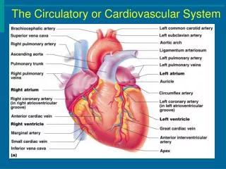
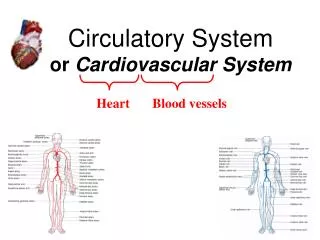
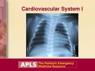
Cardiovascular or Circulatory System I Introduction Structure of Heart Cardiac Cycle Conductive Pathway
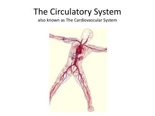
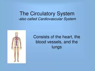
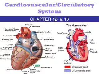
Introduction • Circulatory system = Cardiovascular system • Transportation system of the body • Consists of • Heart • Blood vessels • Blood • Transports • oxygen and nutrients to body cells • carbon dioxide and metabolic materials away from body cells
Heart • A muscular, hollow organ • “Pump” of the body • Weighs about 1 pound • Size of closed fist • Contracts 100,000 times per day • Pumps 2000 gallons of blood daily • Located in mediastinal cavity • Between lungs • Behind sternum • Above diaphragm
Views of the Heart Posterior view Interior on coronal plane Anterior view
Three layers of tissue form the heart • Endocardium • Smooth layer of cells that lines the inside of the heart inside of blood vessels • Allows for smooth flow of blood • Myocardium • Thickest layer • Muscular layer of heart • Pericardium • Double-layered membrane • Covers outside of heart • Thin layer of fluid (pericardial fluid) between the double layer for lubrication
Septum • Muscular wall that separates the heart into a right side and left side • Prevents blood from moving between the right and left sides of the heart • Upper part is called the interatrial septum • Lower part is called the interventricular septum
Heart Chambers • Heart is divided into four chambers • 2 Upper chambers = atria • 2 Lower chambers = ventricles • Right atrium • Receives blood as it returns from the body cells • Right ventricle • Receives blood from the right atrium and pumps the blood into pulmonary artery which carries it to the lungs for oxygen
Left atrium • Receives oxygenated blood from the lungs • Left ventricle • Receives blood from the left atrium and pumps the blood into the aorta for transport to the body cells
CheckPoint 1. What are the names of the heart chambers? a. atria and ventricles b. upper and lower c. left and right.
CheckPoint 2. The muscular layer of the heart is the __________ ? a. endocardium b. myocardium c. pericardium.
CheckPoint 3. The right side of the heart contains oxygenated or deoxygenated blood?
Valves -Located in chambers of heart • Keeps blood flowing in correct direction • Tricuspid valve • Located between the right atrium and right ventricle • Closes when the right ventricle contracts • Pulmonary valve • Located between the right ventricle and the pulmonary artery. • Closes when the right ventricle has finished contracting • Mitral (Bicuspid) valve • Located between the left atrium and the left ventricle • Closes when the left ventricle is contracting • Aortic valve • Located between the left ventricle and the aorta (largest artery in the body) • Closes when the left ventricle is finished contracting
Cardiac Cycle (Heartbeat cycle) Right and Left sides work together in cyclic manner Electrical impulses cause myocardium to contract Cycle consists of a brief period of rest – called diasystole Followed by a period of ventricular contraction – called systole
Process of Contraction At start of the cycle, atria contract and push blood into the ventricles The atria then relax, and blood returning from the body enters the right atrium, while blood returning from the lungs enters the left atrium As atria are filling, systole begins and ventricles contract Right ventricle pushes blood into the pulmonary artery into the lungs, while the left ventricle pushes blood into the aorta and to other parts of the body.
Blood on right side of heart is deoxygenated (low in oxygen, high in carbon dioxide) • Needs to go to the lungs for gas exchange • Blood on left side of heart is oxygenated (high in oxygen, low in carbon dioxide) • Ready for transport to body cells
Blood flow through heart • RIGHT ATRIUM • Receives deoxygenated blood as it returns from the body • Blood enters right atrium from Inferior and Superior Vena Cava (largest veins in the body)
Blood flow through heart (cont.) • RIGHT VENTRICLE • Receives deoxygenated blood from the right atrium via the tricuspid valve • Pushes blood into the pulmonary artery through the pulmonic valve which carries blood to the lungs for oxygen
LEFT ATRIUM Receives oxygenated blood from the lungs via the pulmonary vein
LEFT VENTRICLE • Receives oxygenated blood from the left atrium via the bicuspid or mitral, valve • Pushes blood into the aorta through the aortic valve • Aorta is main artery that sends blood to rest of the body • Largest artery in body
CheckPoint 4. The valve between the right atrium and right ventricle is the __________ ? a. mitral valve b. aortic valve c. tricuspid valve.
CheckPoint 5. The valve between the left atrium and left ventricle is the __________ ? a. mitral valve b. aortic valve c. tricuspid valve.
Video of blood flow through heart • http://www.youtube.com/watch?v=JA0Wb3gc4mE • http://www.youtube.com/watch?NR=1&v=Bfn7vO_BJJY&feature=fvwp • Bill Nye the Science Guy: • http://www.youtube.com/watch?NR=1&v=GbttJ-5do9M&feature=endscreen • http://www.youtube.com/watch?feature=endscreen&v=RiYOuI7iyp8&NR=1
Conduction through the Heart • Electrical impulses starting in the heart cause contraction of the muscles. • Parts of the conduction system • SA Node • AV Node • Bundle of HIS • Purkinje fibers
Sinoatrial Node (SA Node) Group of nerves located in right atrium Known as the “Pacemaker of the heart” Sends electrical impulse that spreads over muscle of atria Atrial muscles contract, then push blood into ventricles. After impulse goes through atria, it reaches the Atrioventricular (AV) node.
Atrioventricular Node (AV Node) Group of nerve cells Located between the atria and ventricles Electrical impulse through septum Nerve fibers in septum called bundle of HIS
Bundle of HIS Nerve fibers in septum Separate into the Right and Left Bundle Branches
Right and Left BUNDLE BRANCHES Carry impulse down through the ventricles Subdivides into a network of nerve fibers in ventricle called Purkinje fibers.
Purkinje Fibers Final conduction pathway Spread to all muscle tissue in the ventricles Cause ventricles to contract
CONDUCTION Occurs approximately every 0.8 seconds. Electrical impulse can be recorded on an EKG Used to detect abnormal activity or disease.
CheckPoint 6. Name the part known as the “pacemaker of the heart”. a. AV node b. SA node c. Purkinje fibers d. Bundle of HIS.
CheckPoint 7. Name the part that runs down the septum of the heart. a. AV node b. SA node c. Purkinje fibers d. Bundle of HIS.
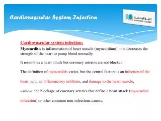
Arrhythmias • Abnormal heart rhythms • Can be Mild to life-threatening • Examples: • Premature atrial contraction • Ventricular fibrillation • Life-threatening ones can be treated with a defibrillator – device that shocks heat to momentarily stop it and allows SA node to regain control • Pacemaker – battery-powered device that monitors heart and emits an electrical impulse to stimulate contraction
Two Pacemakers: SA and AV Nodes SA Node fires 60-80 bpm AV Node fires 40-60 bpm Normal HR 80 bpm Tachycardia >100bpm Bradycardia <60 bpm Review
CheckPoint 8. Name the conduction pathway of the heart in the correct order. a. b. c. d. e.