Download
1 / 22
220 likes | 268 Vues
Learn about the structure, synthesis, and regulation of hemoglobin, along with anemia causes and diagnosis methods. Discover how Erythropoietin affects hemoglobin levels and explore factors influencing native hemoglobin function.

E N D
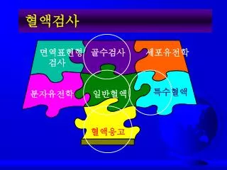
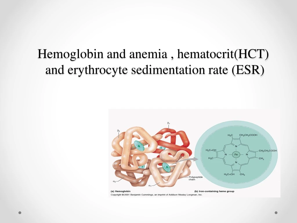
Hemoglobin and anemia , hematocrit(HCT) and erythrocyte sedimentation rate (ESR)
Hemoglobin synthesis • The circulation blood of normal adult contain about 750 mg of hemoglobin and about 7 – 8 gm are degraded daily. • This amount has to be newly synthesized each day because: • The globin part of Hemoglobin can be reutilized only after catabolism into its constituent amino acid • The free hemeis broken down into bile pigment which is excreted. • Iron alone is reutilized in the synthesis of hemoglobin. • The rate at which hemoglobin is synthesized and which red cell are formed are related to : • Oxygen content of the blood • Capacity of the blood to carry oxygen ,which in turn depend on the amount of circulating hemoglobin
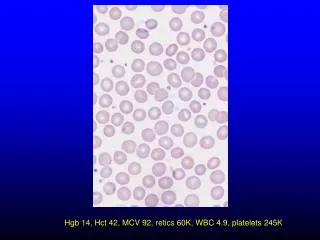
immature RBC Reticulocyte develop and mature in the red Bone marrow and then circulate for about a day in the blood stream before developing into mature red blood cells. Like mature red blood cells, reticulocytes do not have a cell nucleuses They are called reticulocytes because of a reticular network of ribosomal RNA that becomes visible under a microscope with certain stains such as new methylene blue
Regulation of hemoglobin synthesis: Erythropoietin : is formed in kidney in response to decrease oxygen carrying capacity (hypoxia or anemia), in order to stimulate the erythropoiesis or hematopoiesis *anoxia: means a total depletion in the level of oxygen an extreme form of hypoxia or "low oxygen
Tissue hypoxia Kidney secrete erythropoietin into blood Increase erythropoiesis Increase number of RBC Increase oxygen carrying capacity Return to homeostasis when oxygen deliver to kidney , this cause negative feedback inhibition to stop secrete of erythropoietin
Hemoglobin catabolism: • In spleen the reticuloendothelial system thatdestroyed erythrocytes and hemoglobin is released. • Globin is separated from hemeand haematin is formed (the iron oxidized to feeric iron Fe+3) • The porphyrin ring is then opened and the iron is removed with formation of straight chain compundbiliverdin which is converted to bilirubine by reduction • The iron and amino acid of the globin are retained but pyrrole ring are excreted as bilirubin.
The role of some factor affecting on the native of haemoglobin: • Vitamins • cofactors Deficiencies : such as biotin , coenzyme A and pyridoxal phosphate are essential for hemesynthesis . The folic acid deficiencies can cause megaloblastic anemia . • trace metals : only copper and cobalt are known to play a role .(copper is playing a role in the absorption of iron while cobalt is essential constituent of vitamin B12(Cobalamin) ) • ** The causes which lead to deficiency of Vit B12? • Deficiency of cobalt or /and deficiency in intrinsic factor • 4) Glucose -6-phsphatase dehydrogenase (G6PD) deficiency : G6PD is enzyme responsible for the conversion the glucose into pentose phosphate pathway to form 6-phosphogluconate , this pathway provide NADPH which use for produced reduced glutathione and for other reaction such as reduction methaemoglobin.
Deficiency of G6PD lead to Decrease level of NADPH , and this lead to broken of RBC membrane then cause hymolysis
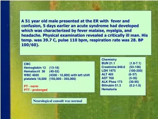
Anemia : Is in general decrease in number of RBC or less normal quantity of hemoglobin in blood leading to decrease oxygen binding capacity . Causes: Genetics Acquired RBC membrane Defect Megaloblastic Anemia Iron-Deficiency Anemia
Iron-deficiency anemia: Deficiency of iron is essentially due to blood loss with failure to replace the iron stores because of : dietary deficiency or increase requirement or defective absorption all of this causes lead to decrease plasma iron . Megaloblastic Anemia: This may be due to deficiency of folic acid or cobalamins (Vit. B12) RBC membrane defects: In this condition there is a defect of the erythrocyte membrane and an abnormally in the sodium pumps.
Low Hb. Indicating : anemia (mention all types of anemia) High Hb. Indicating : polycythemia vera (abnormally increasing of RBC) Normal Hemoglobin conc. : 12 – 18 g/dl
Estimation of blood haemoglobin: Principle: The ferrouse (Iron II) in each hemein RBC is oxidized by ferricyanide to Fe(III)-methaemoglobin . A cynide group is then attached to the iron atom (because it is positively charge) by reaction with KCN to give the brown cyanmethamoglobin (stable)which can be estimated quantitatively
Calculation Hemoglobin Conc. In test =Absorbance of test x 29.4 Hemoglobin Conc. In test = g/dl Normal Haemoglobin conc. : FOR men:14 – 18 g/dl For women : 12_16 g\dl
Erythrocyte sedimentation rate (ESR) • ESR is the rate at which RBC sediment in a period of one hour. It is a common hematology test, and is a non-specific measure of inflammation .
ESR anticoagulated blood is placed in an upright tube, known as a Westergren tube, and the rate at which the RBC fall is measured and reported in mm/h. (millimeter per hr) **inflammation or Anemia changes the proteins in red blood cells causing them to bind to one another in clumps, making them denser than normal red blood cells.
Method: BY Westergren method:When anticoagulatedwhole blood is allowed to stand in a narrow vertical tube the RBCs – under the influence of gravity - settle out from the plasma. (mm/hr).
Average values in healthy men are: <15mm/hr; in healthy females, <20mm. The values are slightly higher in old age, in both genders.
hematocrit HCT The hematocrit is the percentage of blood composed of red blood cells. People with a high volume of plasma (the liquid portion of blood) may be anemic even if their blood count is normal because the blood cells have become diluted. Like hemoglobin, (reduce in HCT indicate for Anemia) a normal hematocrit percentage depends on age and gender. Normal results vary, but in general are as follows: Male: 40.7 - 50.3% Female: 36.1 - 44.3% Interpretation : Low HCT cause of anamemia High HCT cause of Polycythemia Calculation : HCT= Length of RBC colum Total lengethofbloodcolum