
The Impact of 2D and 3D Culture Systems on Osteogenic Differentiation of Human MSCs
E N D
Presentation Transcript
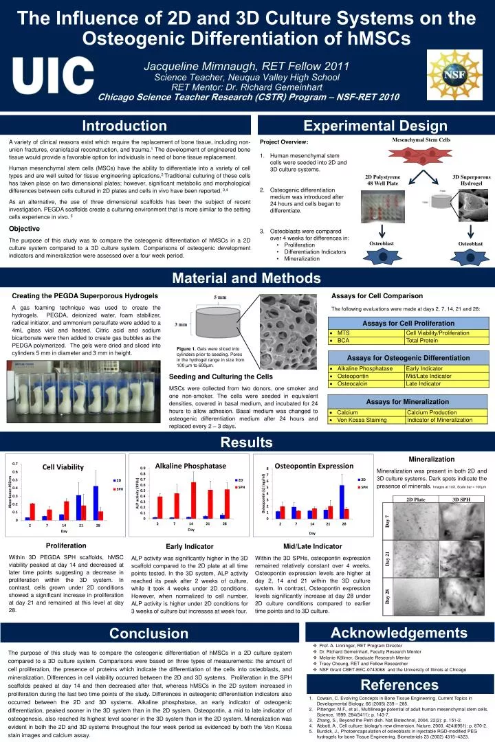
The Influence of 2D and 3D Culture Systems on the Osteogenic Differentiation of hMSCs Jacqueline Mimnaugh, RET Fellow 2011Science Teacher, Neuqua Valley High SchoolRET Mentor: Dr. Richard GemeinhartChicago Science Teacher Research (CSTR) Program – NSF-RET 2010 Introduction Experimental Design A variety of clinical reasons exist which require the replacement of bone tissue, including non-union fractures, craniofacial reconstruction, and trauma.1 The development of engineered bone tissue would provide a favorable option for individuals in need of bone tissue replacement. Human mesenchymal stem cells (MSCs) have the ability to differentiate into a variety of cell types and are well suited for tissue engineering aplications.2 Traditional culturing of these cells has taken place on two dimensional plates; however, significant metabolic and morphological differences between cells cultured in 2D plates and cells in vivo have been reported. 3,4 As an alternative, the use of three dimensional scaffolds has been the subject of recent investigation. PEGDA scaffolds create a culturing environment that is more similar to the setting cells experience in vivo. 5 Objective The purpose of this study was to compare the osteogenic differentiation of hMSCs in a 2D culture system compared to a 3D culture system. Comparisons of osteogenic development indicators and mineralization were assessed over a four week period. • Project Overview: • Human mesenchymal stem cells were seeded into 2D and 3D culture systems. • Osteogenic differentiation medium was introduced after 24 hours and cells began to differentiate. • Osteoblasts were compared over 4 weeks for differences in: • Proliferation • Differentiation Indicators • Mineralization Material and Methods Creating the PEGDA Superporous Hydrogels A gas foaming technique was used to create the hydrogels. PEGDA, deionized water, foam stabilizer, radical initiator, and ammonium persulfate were added to a 4mL glass vial and heated. Citric acid and sodium bicarbonate were then added to create gas bubbles as the PEDGA polymerized. The gels were dried and sliced into cylinders 5 mm in diameter and 3 mm in height. Assays for Cell Comparison The following evaluations were made at days 2, 7, 14, 21 and 28: Figure 1. Gels were sliced into cylinders prior to seeding. Pores in the hydrogel range in size from 100 µm to 600µm. Seeding and Culturing the Cells MSCs were collected from two donors, one smoker and one non-smoker. The cells were seeded in equivalent densities, covered in basal medium, and incubated for 24 hours to allow adhesion. Basal medium was changed to osteogenic differentiation medium after 24 hours and replaced every 2 – 3 days. Results Mineralization Mineralization was present in both 2D and 3D culture systems. Dark spots indicate the presence of minerals. Images at 10X, Scale bar = 100µm Proliferation Within 3D PEGDA SPH scaffolds, hMSC viability peaked at day 14 and decreased at later time points suggesting a decrease in proliferation within the 3D system. In contrast, cells grown under 2D conditions showed a significant increase in proliferation at day 21 and remained at this level at day 28. Mid/Late Indicator Within the 3D SPHs, osteopontin expression remained relatively constant over 4 weeks. Osteopontin expression levels are higher at day 2, 14 and 21 within the 3D culture system. In contrast, Osteopontin expression levels significantly increase at day 28 under 2D culture conditions compared to earlier time points and to 3D culture. Early Indicator ALP activity was significantly higher in the 3D scaffold compared to the 2D plate at all time points tested. In the 3D system, ALP activity reached its peak after 2 weeks of culture, while it took 4 weeks under 2D conditions. However, when normalized to cell number, ALP activity is higher under 2D conditions for 3 weeks of culture but increases at week four. Acknowledgements Conclusion • Prof. A. Linninger, RET Program Director • Dr. Richard Gemeinhart, Faculty Research Mentor • Melanie Köllmer, Graduate Research Mentor • Tracy Choung, RET and Fellow Researcher • NSF Grant CBET-EEC-0743068 and the University of Illinois at Chicago The purpose of this study was to compare the osteogenic differentiation of hMSCs in a 2D culture system compared to a 3D culture system. Comparisons were based on three types of measurements: the amount of cell proliferation, the presence of proteins which indicate the differentiation of the cells into osteoblasts, and mineralization. Differences in cell viability occurred between the 2D and 3D systems. Proliferation in the SPH scaffolds peaked at day 14 and then decreased after that, whereas hMSCs in the 2D system increased in proliferation during the last two time points of the study. Differences in osteogenic differentiation indicators also occurred between the 2D and 3D systems. Alkaline phosphatase, an early indicator of osteogenic differentiation, peaked sooner in the 3D system than in the 2D system. Osteopontin, a mid to late indicator of osteogenesis, also reached its highest level sooner in the 3D system than in the 2D system. Mineralization was evident in both the 2D and 3D systems throughout the four week period as evidenced by both the Von Kossa stain images and calcium assay. References Cowain, C. Evolving Concepts in Bone Tissue Engineering. Current Topics in Developmental Biology, 66 (2005) 239 – 285. Pittenger, M.F., et al., Multilineage potential of adult human mesenchymal stem cells. Science, 1999. 284(5411): p. 143-7. Zhang, S., Beyond the Petri dish. Nat Biotechnol, 2004. 22(2): p. 151-2. Abbott, A., Cell culture: biology's new dimension. Nature, 2003. 424(6951): p. 870-2. Burdick, J., Photoencapsulation of osteoblasts in injectable RGD-modified PEG hydrogels for bone Tissue Engineering. Biomaterials 23 (2002) 4315–4323.