Module G: Oxygen Transport
Module G: Oxygen Transport. Topics to Cover. Oxygen Transport Oxygen Dissociation Curve Oxygen Transport Studies Tissue Hypoxia Cyanosis Polycythemia. Oxygen Transport. Oxygen is carried from the lungs to the tissues in two different fashions:

Module G: Oxygen Transport
E N D
Presentation Transcript
Topics to Cover • Oxygen Transport • Oxygen Dissociation Curve • Oxygen Transport Studies • Tissue Hypoxia • Cyanosis • Polycythemia
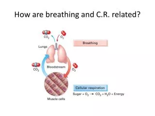
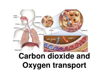
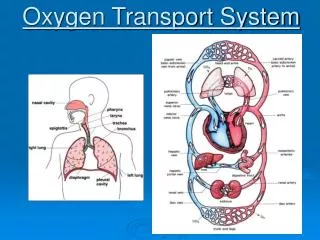
Oxygen Transport • Oxygen is carried from the lungs to the tissues in two different fashions: • Oxygen Dissolved in the blood plasma (PO2) • Oxygen Combined with Hemoglobin (SO2)
Dissolved Oxygen • As oxygen diffuses across the A-C membrane, it dissolves into the plasma and remains in that fashion until it reaches the tissue. • Expressed as a partial pressure - PO2 • Normal arterial level = 80-100 mm hg • Normal venous level = 35 – 45 mm hg • The quantity of oxygen dissolves is a function of Henry’s law. • At 37° C 0.003 ml of oxygen will dissolve in 100 ml of blood. (Henry’s Law) • At 100 mm hg PaO2, the amount of dissolved oxygen is 0.3 ml O2/100 ml blood (0.3 vol%).
Combined Oxygen • Dissolved oxygen is inadequate for metabolic needs. • Need a substance which will bind to oxygen and carry it, BUT will release it when needed. • Voila! Hemoglobin! • Miracle #2…As we’ll see later, hemoglobin also carries carbon dioxide and is a key buffer of [H+].
Hemoglobin • Each RBC contains approximately 280 million molecules of hemoglobin • Adult Hemoglobin (Hb A) consists of • 4 Heme Groups – Primarily iron in the Fe+2 (ferrous) state. • 4 Globulin – 4 amino acid chains (2 a & 2 b) • One heme group binds with one of the amino acid chains and oxygen binds with the iron in the heme group in a reversible way. O2 + Fe+2« FeO2 (Oxyhemoglobin) FERROUS
achains bchains Heme Units
Hemoglobin & the RBC • “Normal” hemoglobin level • 15 g/dl (men) & 13-14 g/dl (women) - Malley • 15 + 1.5 g/dl (men) & 13.5 + 1.5 g/dl (women) – Egan • 15 + 2 (men) & 14 + 2 (women) - Easy • Why do women have less? • The hemoglobin molecule resides in the erythrocyte and is responsible for giving the blood its red color. • Hundreds of hemoglobin variants. • Normal: A, A2, F • Abnormal: S, H
The affinity of hemoglobin for oxygen increases with each oxygen molecule attached • “All or Nothing”
Oxygen Saturation • Saturation = Sites Filled Total Sites Available • Example: • 80 sites filled = 80% saturation 100 sites available • Oxygen Saturation only talks about how much hemoglobin is saturated, NOT how much hemoglobin is present.
Combined Oxygen • Each gram of Hemoglobin combines with 1.34 mL of oxygen. • O2 bound to Hb = 1.34 mL O2 x 15 g Hb = 20.1 vol% O2
Quantity of Oxygen Bound to Hemoglobin • Not all hemoglobin molecules are bound with oxygen. • Normal saturation • Arterial (SaO2) – 97% • Venous (SO2) – 75% • Some “desaturated” hemoglobin exists because of normal physiologic shunts: • Mixing of poorly saturated venous blood with arterial blood • Thebesian, bronchial, and pleural veins • Intrapulmonary shunts (perfused alveoli that are not ventilated) • 20.1 vol% O2 x 0.97 = 19.5 vol% O2 • Hemoglobin not bound with oxygen is called reduced hemoglobin.
Oxygen Content • The total amount of oxygen in 100 mL of blood is the sum of the dissolved oxygen & the oxygen bound to hemoglobin. • Arterial Content • CaO2 = (Hb x 1.34 x SaO2) + (PaO2 x .003) • Venous Content • CO2 = (Hb x 1.34 x SO2) + (PO2 x .003) • Capillary Content • CćO2 = (Hb x 1.34 x SćO2) + (PćO2 x .003) • SćO2 = Ideal Saturation = 100% • PćO2 = Ideal Partial Pressure = PAO2
Oxygen Dissociation Curve • The relationship between the partial pressure of oxygen and the saturation of oxygen is not linear. • 2 lines
Steep Portion of Curve • “Dissociation Portion” of curve. • Between 10 and 60 mm Hg. • Small increases in PO2 yield large increases in SO2. • At the tissue capillary, blood comes in contact with reduced tissue PO2 and oxygen diffuses from the capillary to the tissue. As the PO2 falls, oxygen bound to the hemoglobin (SO2) is released.
Flat Portion of Curve • “Association Portion” of curve. • Greater than 60 mm Hg. • Large increases in PO2 yield small increases in SO2. • At the pulmonary capillary, blood comes in contact with increased alveolar PO2 and oxygen diffuses from the alveolus to the capillary. As the PO2 rises, oxygen binds with the hemoglobin (increasing SO2). • Very little rise in oxygen saturation above 100 mm Hg of PaO2.
Key Points to Remember • The amount of oxygen that is saturated on the hemoglobin (SO2) is dependent on the amount dissolved (PO2). • Between a PaO2 of 60 and 100 mm Hg, the curve is flat. • This means that small changes in PaO2 can occur without a big drop in saturation and oxygen content. • At the tissues, where the PO2 is about 40 mm Hg, the curve is steep. • This means that small changes in PO2 will result in a large amount of oxygen being unloaded.
P50 • The degree to which oxygen is attracted to hemoglobin can be assessed by evaluating at what PO2 there is a 50% SO2(P50). • Normal P50 value is 27 mm Hg • As P50 increases/decreases, we say the “curve has shifted”. • P50 less than 27: Shift to the left. • P50 greater than 27: Shift to the right.
Leftward Shift of the OHDC • Left Shift. • Increased oxygen affinity for hemoglobin. • This is the expected shift at the lungs (Left-Lungs). • For any given PO2, SO2 will be higher. • Decreasing P50. • LOW THINGS CAUSE A LEFT SHIFT • ¯H+ concentration (pH, alkalosis) • ¯Temperature (Hypothermia) • ¯ Carbon Dioxide (PCO2, Hypocarbia) • ¯ 2,3 DPG
Rightward Shift of the OHDC • Right Shift. • Decreased oxygen affinity for hemoglobin. • This is the expected shift at the tissues. • For any given PO2, SO2 will be lower. • Increasing P50. • ELEVATED THINGS CAUSE A RIGHT SHIFT (Up-Right) • H+ concentration (¯ pH, acidosis) • Temperature (Hyperthermia) • Carbon Dioxide (PCO2, Hypercarbia) • 2,3 DPG
2,3 DPG • 2,3 DPG is an organic phosphate normally found in the RBC that has a tendency to bind with Hemoglobin and thereby decrease the affinity of Hemoglobin for oxygen. • It promotes a rightward shift and enhances oxygen unloading at the tissues. This shift is longer in duration than that due to [H+], PCO2 or temperature. • A doubling of DPG will result in a 10 torr increase in P50.
2,3 DPG • The levels increase with • Cellular hypoxia. • Anemia • Hypoxemia secondary to COPD • Congenital Heart Disease • Ascent to high altitudes • The levels decrease with • Septic Shock • Acidemia • Stored blood • No DPG after 2 weeks of storage.
Bohr Effect • The effect of CO2 on the OHDC is known as the Bohr Effect. • (OH – Bohr) • High PCO2 levels and low pH decrease affinity of hemoglobin for oxygen (a right-ward shift). • This occurs at the tissues where a high level of PCO2 and acidemia contribute to the unloading of oxygen.
Oxygen Transport • The volume of oxygen leaving the left ventricle each minute. • CaO2 x CO x 10 = 20 ml O2 x 5 L/min x 10 • Make sure to convert CO to L/min! • Normal value is 1,000 mL O2 / min • Decreased oxygen delivery occurs when there is: • A decreased blood oxygenation • Decreased hemoglobin concentration • Decreased cardiac output.
Arterial-Venous Oxygen Content Difference • C(a-)O2 • CaO2 – CO2 • The venous blood is “mixed venous” blood obtained from the pulmonary artery via a pulmonary artery catheter. • Normal CaO2: 20 vol% • Normal CO2: 15 vol% • Normal CaO2 – CO2: 5 vol% • Decreased with: • Increased CO • Certain Poisons • Hypothermia
Oxygen Extraction Ratio • Def: The amount of oxygen extracted by the peripheral tissues divided by the amount of oxygen delivered to the peripheral cells. • aka: Oxygen coefficient ratio & Oxygen utilization ratio • O2ER = (CaO2-CO2)/CaO2 • Normal is 25% • Increased with: • Decreased CO • Increased VO2 • Exercise • Seizures • Shivering • Hyperthermia • Anemia • Low PaO2 • Decreased with: • Increased Cardiac Output • Skeletal Muscle Relaxation • Peripheral Shunting • Certain Poisons • Hypothermia • Increased Hemoglobin • Increased PaO2 .
Pulmonary Shunting • PERFUSION WITHOUT VENTILATION. • Pulmonary shunt is that portion of the cardiac output that enters the left side of the heart without coming in contact with an alveolus. • “True” Shunt – No contact • Anatomic shunts (Thebesian, Pleural, Bronchial) • Cardiac anomalies • “Shunt-Like” (Relative) Shunt • Some ventilation, but not enough to allow for complete equilibration between alveolar gas and perfusion. • Shunts are refractory to oxygen therapy. • Delivery of oxygen therapy will NOT help (at least to the expected degree).
Venous Admixture • The mixing of oxygenated blood with “contaminated” deoxygenated blood resulting in a reduction in: • PaO2 • SaO2
Alveolar-arterial Gradient • PAO2-PaO2 • More on this in today’s 1060 class.
Shunt Equation • So just how much blood is shunted? • Where CćO2 = (Hb x 1.34) + (PAO2* .003) • You will need • PBARO – Barometric Pressure • PaO2 – Arterial Partial Pressure of Oxygen • PaCO2– Arterial Partial Pressure of Carbon Dioxide • PO2– Venous Partial Pressure of Oxygen • Hb – Hemoglobin concentration • PAO2 – Alveolar Partial Pressure of Oxygen • FIO2 – Fractional Concentration of Inspired Oxygen
Shunt Equation Example You obtain the following data on you patient. What is the percent shunt present? pH: 7.25 PaCO2: 28 torr PaO2: 68 torr SaO2: 91% PB: 749 PO2: 38 torr SO2: 72 % Hb: 13 gm% FIO2: .90 • Steps: • Calculate CaO2 • Calculate CO2 • Calculate PAO2 • Calculate CćO2 • Plug in numbers & solve!
Shunt – Clinical Significance • Normal Shunt: 3 to 5% • Shunts above 15% are associated with significant hypoxemia.
Tissue Hypoxia • Hypoxemia: Reduced oxygen in blood (PaO2). • Hypoxia: Reduced oxygen at the tissue. • Four types: • Hypoxic (Hypoxemic) Hypoxia • Anemic Hypoxia • Circulatory Hypoxia • Histotoxic Hypoxia
Hypoxic Hypoxia • Tissues have inadequate oxygen levels because there is inadequate levels in the blood (reduced PaO2). • Rule out causes of hypoxemia • Low Alveolar Oxygen (reduced PAO2) • Altitude • Hypoventilation (increased PACO2) • Breathing of gas mixtures less than 21% • Diffusion Impairment • Intrapulmonary Shunt • Ventilation/Perfusion Mismatch
Anemic Hypoxia • Tissues have inadequate oxygen levels secondary to reduced oxygen carrying capacity. • No hypoxemia! • Caused by • Reduced Hemoglobin • Defective Hemoglobin • Carboxyhemoglobin • Methemoglobin • Sulfahemoglobin • Sickle Cell Anemia (HbS)
Carboxyhemoglobin • Carbon Monoxide has 245 times the affinity for hemoglobin as oxygen. • Causes a leftward shift (increased affinity of Oxygen) • Normal level is less than 3% • 10% is common with smokers. • Toxic at 20%; Lethal at 50%. • Suspect incomplete combustion if level is elevated. • Furnace, space heater • Treatment is 100% oxygen • Reduces half-life of HbCO from 5 hours to 20 minutes. • Hyperbaric treatment remains controversial.
Methemoglobin • A normal variant of adult hemoglobin. • Ferrous to Ferric (loses an electron, Fe+3) • Normal levels are less than 1%. • Usually associated with excessive nitrate ingestion. • Amyl Nitrate • Nitroglycerine • Topical anesthetics • Treatment with methylene blue
Sulfhemoglobin • Sulfhemoglobin results from the union of hemoglobin with medications such as sulfonamides (antibiotics including Bactrim & Septra). • The resultant form of hemoglobin is unable to transport oxygen, and is untreatable. • The only treatment is to wait until the affected red blood cells are destroyed as part of their normal life cycle.
Hemoglobin S • Hemoglobin S is a abnormal variant of Hemoglobin A where one of the 146 amino acids in the beta chain is altered. • Inherited disorder from both parents. • 1% of African Americans. • Recurrent painful episodes (Crisis) occur as sickled cells become obstructed. • Typical “anemic” symptoms. • Can lead to infections and stroke.
Circulatory Hypoxia • Tissues have inadequate oxygen levels secondary to reduced oxygen delivery. • Most significant cause is reduced cardiac output or blood pressure abnormalities.
Histotoxic Hypoxia • Tissues have inadequate oxygen levels secondary to inability of tissue to use oxygen for metabolism. • Cyanide poisoning.
Cyanosis • A clinical condition manifested by a bluish discoloration of the mucous membranes or nail beds. • Peripheral vs. Central • Present when 5 g/dl of hemoglobin is desaturated. • This usually correlates with a SaO2 below 85%. • ANEMIC PATIENTS WILL NEVER BE CYANOTIC!
Polycythemia • Response to chronic hypoxemia is the release of erythropoietin and stimulation of the bone marrow to produce more RBC and hemoglobin. • Increased hemoglobin allows for greater carrying capacity BUT also results in increased viscosity of the blood. • Increased viscosity increases work of the heart.
Fetal Hemoglobin (HbF) • Fetal hemoglobin (hemoglobin F) is the main hemoglobin that transports oxygen around the body of the developing baby during the last 7 months of pregnancy. • It has a greater affinity for oxygen than Hemoglobin A (P50 of 20 mm Hg). • At about 30 weeks gestation, the fetus begins to make increasing amounts of hemoglobin A. • Hemoglobin F does not turn into hemoglobin A. • As they grow babies automatically turn off the production of hemoglobin F (usually complete by one year). Failure to stop Hemoglobin F production is found in certain beta thalassemias. Possible link to SIDS.