Download
1 / 19
240 likes | 625 Vues
Calcium and phosphate homeostasis and hyperparathyroidism. Charles Hand. vitamin D 2 (diet). Synthesis of active vitamin D. 10% ,. 90% ,. Bile Salts. calcidiol. 25-hydroxylase. 1,25(OH) 2 D 3 calcitriol. 1 a -hydroxylase. Tightly regulated.

E N D
Calcium and phosphate homeostasis and hyperparathyroidism Charles Hand
vitamin D2 (diet) Synthesis of active vitamin D 10%, 90%, Bile Salts calcidiol 25-hydroxylase 1,25(OH)2D3 calcitriol 1a-hydroxylase Tightly regulated
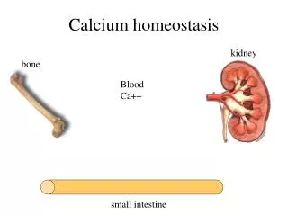
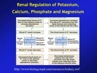
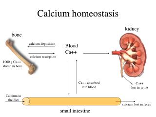
Low calcium is sensed by the parathyroid gland • PTH is released and levels of PTH go up • PTH has 3 effects: • Increase activity of 1aHydroxylase • Increase bone resorption: leads to calcium and phosphate release • Increase Calcium uptake at kidney, and decrease phosphate re uptake • In the above example on the slide, calcium levels are low in the blood, but phosphate levels will be normal. As bone resorption occurs, calcium and phosphate levels will rise. This will correct the calcium levels but increase the phosphate levels. Thus, phosphate reuptake is decreased at the kidney in order to counteract this situation. • 1aHydroxylase catalyses the conversion of calcidiol to calcitriol, the active form of vitamin D3. • Calcitriol then has 4 main effects: • Increase bone resorption • Increase calcium and phosphate reuptake at the kidney • Increase calcium and phosphate uptake in the gut • Negatively feeds back to the parathyroidgland • Bone resorption triggers FGF23 release which prevents phosphate levels becoming too high • Calcium levels rise which leads to: • Negative feedback to the parathyroid • Calcitonin release to decrease calcium and phosphate reabsorption at the kidney Calcium (PO4) homeostasis PTH bone resorption Ca2+release KIDNEY BONE GUT 25(OH)D3 1-OHase 1,25(OH)2D3 Ca2+ & PO4 absorption bone resorption Ca2+ release Ca2+ reabsorption PO4 reabsorption FGF23 Ca2+ High Ca2+ thyroid gland/calcitonin low Ca2+ PARATHYROIDS
Phosphate homeostasis PARATHYROIDS PTH - - low PO4 + KIDNEY Klotho GUT calcidiol - FGF23 1-OHase BONE calcitriol ↑ PO4 absorption ↑ PO4 release PO4 reabsorption PO4 feedback -
Hyperparathyroidism • What is it? • Over activity of parathyroid gland causing excess parathyroid hormone (PTH) production • What are the consequences? • Damage to bone • What are the types? • Primary • Secondary • Tertiary
Primary • What is it? • Over activity of the parathyroid glands themselves. • Causes? • Adenoma • Hyperplasia • Carcinoma (rare)
Secondary • What is it? • Normal response to low calcium levels • Causes? • Vit D deficiency main cause • Chronic renal failure • Consequences? • Renal osteodystrophy
Tertiary • What is it? • Seen in chronic 2ndary hyperparathyroidism • Hyperplasia of parathyroid glands • Become unresponsive to changes in Ca2+ • Gets stuck on high production mode • Who gets it? • Patients with chronic renal failure
Exam Q (ESA 3 2011) Question 8 A 54 year woman presents to A&E complaining of severe back pain that radiates into the groin. You suspect she has a kidney stone. A. Give THREE pieces if information that you could obtain from taking a clinical history of a patient that would indicate why a kidney stone had formed.
Exam technique • Make sure with each new question you highlight the key points and any traps you are likely to fall into: • Patient demographics, age, sex. This gives you potentially a big clue • Describe, list, explain. Make sure you do what it asks • Clinical history etc. If it says clinical history, you can’t give information you would gain from an examination • Give THREE. If it says three, make sure you give three! • Never leave a question blank. If you don’t know keep reading ahead, often you will find a clue later in the question. If you still don’t know, just guess! Everyone else may have found that question hard and the marks be brought down, so don’t despair!
A. Answers • Dehydration • Gout, IBD (Crohn’s) • High protein diet • Recurrent infections • Familial
B. Blood tests show that this patient has hypercalcaemia. (i) Why is she likely to have developed a kidney stone? (ii) Name TWO types of stone that are likely to be visible on a plain X-ray film. The stone is located in the abdominal ureter. iii) Where would you expect to see it on a plain X-ray film?
B. Answers • i) • As calcium increases, amount of oxalate in bloodstream for absorption increases. Oxalate then excreted more into the urine. Causes calcium oxalate precipitation. Most common type. • ii) • Calcium phosphate • Calcium oxalate • iii) • PUJ • VUJ • Pelvic brim
C. Further tests demonstrate high parathyroid hormone levels and a single enlarged parathyroid gland. She has primary hyperparathyroidism. • Apart from parathyroid hormone name ONE other hormone which you would expect to be elevated in this patient. • (ii) Explain why she has hypercalcaemia.
C. Answers • i) Calcitriol. Calcitonin? • ii) I’ll this one to you to work out from the diagrams/ text at the beginning of this presentation.
D. Her parathyroid gland is enlarged due to hyperplasia. How does hyperplasia differ from hypertrophy?
D. Answers • Hypertrophy: Enlargement of organ or tissue due to increase in cell size • Hyperplasia: Enlargement of an organ or tissue due to an increase in number of cells
E. Cell division is involved in hyperplasia. Describe the cell cycle, including the four stages of cell division, and indicate where the two checkpoints are located. A diagram may be used.