Download
1 / 24
350 likes | 1.86k Vues
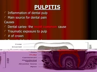
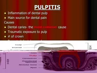
PULPITIS Inflammation of dental pulp Main source for dental pain Causes Dental caries- the most common cause Traumatic exposure to pulp # of crown Repeated dental procedure. Pathogenesis Any causes above Exposure to pulp Invasion by bacteria- Streptococus Inflammation of pulp.

E N D
PULPITIS • Inflammation of dental pulp • Main source for dental pain Causes • Dental caries- the most common cause • Traumatic exposure to pulp • # of crown • Repeated dental procedure
Pathogenesis Any causes above Exposure to pulp Invasion by bacteria- Streptococus Inflammation of pulp
Types • Acute (rapid, severe onset, short duration) • Open – communication between pulp cavity & oral cavity) • closed • Chronic (slow , long duration, mild pain) • Open • Closed
Acute-ClosedMicro organism- virulent & large no. Clinical features • Early stage - Hypersensitivity to hot & cold • Later more persistent • Pain- sharp, severe & stabbing • Sometimes not localized • Tender • Tooth discoloration • Swelling of gum
Acute-Open • Common • Acute exposure with micro organisms • Occurs at late stage of caries • Abscess formed drain out of cavity
Clinical features • Hypersensitivity hot & cold in early stage • Less pain • Slight tender
Chronic-closed • No communication B/W pulp & oral cavity • Pulp tissue destruction at the site of micro organism entry • Infection remains localized for long time with remaing pulp tissue intact or destruction occurs slowly Clinical features • Hypersensitivity • Early stage- to hot n cold • Late stage- only to hot but relieved by cold
Chronic-open • Usually occurs on widely opened cavity • Pulp is destroyed & replaced by granulation tissue & become epithelialised to form polyp • PAINLESS
Diagnosis Test of healthy pulp • Tapping of tooth directly • Sensitivity if present indicates the spread of inflammation to surrounding tissue • hot & cold sensitivity • If pain persists even after stimulus removal or • Pain persists spontaneously Pulp may not be healthy to save
Test of pulp- dead or alive • Electric pulp tester • It helps to recognize the pulp whether it’s alive or dead but not healthiness • If person feels the electric charge delivered to the tooth the pulp is alive
Treatment • If pulp viable just remove irritant n healed itself • Removal of caries n restoration by filling • If pulp dead • RCT • Tooth extraction • antibiotic is given- penicillin in acute cases
Root canal treatment • Root canal treatment is the process where the dentist removes the pulp( i.e. pulp extirpation) from an infected tooth and replaces it with dental filling
Indications for RCT • Dental caries : • an untreated cavityis a common cause of pulp infection. • the reduced blood supply also limits the pulp’s ability to heal itself • Pulpitis • the pulp can become damaged from trauma or facture • Abscess • once the pulp become infected, the infection can spread to the bone around the tooth forming abscess if the rct is not done,the tooth may have to be extracted • Repeated dental procedures on tooth
Procedure • First an opening is made through the occlusal area of posterior teeth and lingual surface of anterior teeth • After the diseased pulp is removed, the pulp chamber & root canal is enlarged & shaped in preparation for being filled • If more than one visit is needed, a temporary filling is placed in the opening of the tooth • The temporary filling is removed and the pulp chamber & root canal permanent restoration is done • In final step, a crown is usually placed over the tooth to restore its natural shiny apperance
all RCT procedures are done by isolating the tooth with a rubber dam to provide saliva-free environment. • RCT may be done in single or multiple visits depending on complexity of the tooth • X-rays are taken to determine the length of the root & to monitor the various aspects of treatment
Filling • Temporary filling material - Zinc oxide or zinc phosphate • Permanent – Gutta Percha