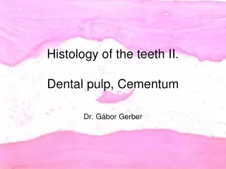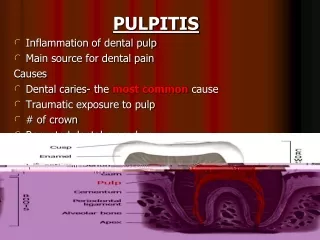
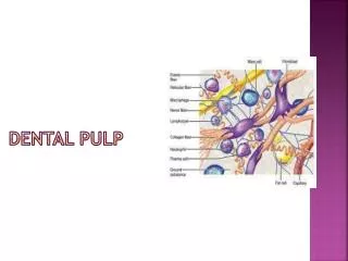
Dental Pulp
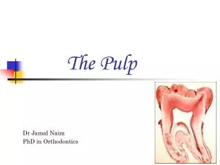
Dental Pulp. Dental Pulp as a connective tissue. Dental pulp is connective tissue uniquely situated within the rigid encasement of mineralized dentin; Although dental pulp shares many properties with other connective tissues of the body;

Dental Pulp
E N D
Presentation Transcript
Dental Pulp as a connective tissue Dental pulp is connective tissue uniquely situated within the rigid encasement of mineralized dentin; Although dental pulp shares many properties with other connective tissues of the body; The peculiar location of dental pulp imposes several special characteristics on it.
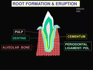
Origin of the dental pulp • The pulp is derived from the mesenchymeandectomesenchyme; • Thepulpis formed from the second element of the tooth germ – dentalpapilla; • The most important developmental events are those guiding epithelial-ectomesenchymal interactions, which involve a molecular crosstolk between the ectoderm and ectomesenchyme, two tissues that have a different origine.
Budstage • As the dental lamina continues to grow and thicken to form a bud, cells of the ectomesenchyme proliferate and condense to form the dental papilla; • At this stage, the inductive or tooth-formating potential is transferred from the dental epithelium to the dental papilla.
Capstage At this stage, the tooth bud assumes the shape of a cap that surrounded the dental papilla; Anotherlayer of mesenchymalcells, calledthedentalfollicle, thatseparatesthetoothorganpapillafromtheotherconnectivetissues of thejaws; The enamel knot expresses a unique set of signaling molecules that influence both the shape of the crown and the development of the dental papilla.
Earlybellstage Thereciprocalexchange of molecularinformationbetweenthedentalorgananddentalpapillainfluencestheimportanteventsthatlead to celldifferentiationatthelatebellstage.
Dentalpapilla • The cardiovascular system originated from cells termed angioblasts, which arise from angiogenic clusters from the visceral mesoderm; • As these cells separate into clusters, the outer cells organize into a series of elongating tubes; • The inner cells become blood cells.
Vascularisation • Vascularisation of the developing pulp starts during the bell stages, with small branches from the principal vascular trunks of the jaw entering the base of the papilla. • The principal vessels enlarge and run through the pulp towards the cuspal regions. • Here they give of numerous small branches, which form a bed of venules, arterioles and capillaries in the sub odontoblastic and odontoblastic layers.
Latebellstage • The most peripheral cells of the dental papilla enlarge and become organized along the basement membrane at the tooth`s epithelial-mesenchymal interface; • Thesenewlydifferentiatedcellsarecalledodontoblasts; • They are the cells that are responsible for the synthesis and secretion of dentin matrix; • Atthistime, dentalpapillaistermedthedentalpulp.
PulpodentinComplex There is a great deal of evidence that dentin and the pulp are functionally coupled and hence integrated as a tissue; Developmentally, pulpalcellsproducedentin, nerves, andbloodvessels; Although dentin and pulp have different structures and compositions, once formed they react to stimuli as a functional unit.
Pulphystogenesis • Beginning of the pulpalhistogenesis is bud stage of tooth; • The end of this process is the completion of the tooth root; • Throughout this period the dental pulp is in its embryonic stage. As more and more dentin is deposited, the pulp chamber narrows and finally forms a canal containing blood vessels and nerves of the tooth.
In all these periods the dental pulp is in its embryonic stage The end of this process is the completion of the tooth root
Embryonicpulp • Incomplete root development.
Periods in the life of the pulp • Embryonic; • Functional; • Senile.
General Propertis of ConnectiveTissue • Connectivetissueisthesupportingtissueswidelydistributedthroughoutthebody; • Themajorconstituent of connectivetissueis: • Extracellular matrix, which is mainly composed of fibrillar protein and ground substance; • Cells – fibroblasts are the principal cells in connective tissue; • Other cellular elements include blood-derived defensive cells, such as macrophages, whose primary function is to cope with infection.
Pulpas a ConnectiveTissues Loose Connective tissue In the central core of the pulp, the basic components are arranged in a manner similar to that found in other loose connective tissues.
Fibriller proteins Compare collagen shown in blue with red side thingies, with elastin shown in yellow.The curly protein shape of elastin proteins and weird cross links going every which way, allow elastin to spring back after stretching, whereas collagen doesn't give so much. • There are two types of fibrillar proteins: • Collagen; • Elastin; • Collagen is the more abundant type and its main component of collagen fibers, which confer strength of the tissue; • Elastinprovideelasticity to thetissue; • Inthepulp, elasticfibersaredistributedonlyinthewalls of thelargerbloodvessels.
fibrillar proteins collagen fibril
The blue diagonal line is a single tropocollagen (pre-collagen unit). They combine into the triple helix (three stranded rope) of a collagen molecule (lower middle). The yellow snaky one is elastin - like a rubber band. The thin cobweb-like fibers are reticulin - immature collagen found mostly in embryos.
Ground substance The proteoglycans are composed of glycosaminoglycan (GAG) chains • Itisresponsibleprimarilyfortheviscoelasticityandfiltrationfunction of connectivetissue; • Itismainlycomposed of: • Proteoglycans, whichconsist of proteincoreandpolysaccharidesidechainscalledglycosaminoglycans; • Adhesiveglycoproteinssuchasfibronectin, whichprimarilyfunction to mediatecell-matrixinteraction.
Illustration of a proteoglycan molecule inside extracellular matrix. Chondroitin sulfate proteoglycans (CSPGs)
Types of connectivetissue Loose connective tissue dense irregular connective tissue Looseconnectivetissue, whichisrichingroundsubstanceandcontainsrelativelyfewerfibers; Denseconnectivetissue, whichischaracterizedby a clearpredominance of collagenfibersandfewercells; Dentalpulpisclassifiedas a looseconnectivetissue.
Themajorfunction of connectivetissueis: • To providematrixthatbindscellsandorgans; • To givessupport to thebody; • Itisalsoresponsibleforvariousactivitiesthatinitiateandorchestratereactions to pathogenicinvasion, andthusitsurvesasanessentialsite of hostdefense; • Italsohas a remarkablecapacity to repairdamagedtissueintheform of scarring.
The First function - To provide matrix that binds cells and organs
StructuralOrganization of thePulp • The pulp consists of coronal and root pulp; • Coronalpulpislargerandcontainsmanymoreelementsthanrootpulp; • Rootpulpactsas a conductingtube to carryblood to andfromthecoronalarea to theapicalcanal; • Bothpulpareascontainthesameelements, althoughthecells, fibers, bloodvessels, andnervesaremorenumerousincoronalpulp.
Histology of Pulp • Peripheral area of pulp is highly organized; • Central pulp - parietal layer or pulp core – pulpal to cell-rich zone.
Architecture of the Pulp • (1) dentin-forming odontoblasts (A). • (2) reticular (Korff's) fibers (B) pass from the central pulp region, across the cell-free zone and between the odontoblasts, their distal ends incorporated into the matrix of the dentin layer; • Numerous capillaries (C) and nerves (D) are also found in this zone; • Just under the cell-free zone is the cell-rich zone (3) containing numerous fibroblasts (E) - the predominant cell type of pulp; • Fibroblasts of the pulp have demonstrated the ability to form as well as degrade collagen; • Perivascular cells (undifferentiated mesenchymal cells) are present in the pulp and can give rise to odontoblasts, fibroblasts or macrophages. • Since odontoblasts themselves are incapable of cell division, any dental procedure that relies on the formation of new dentin (F) after destruction of odontoblasts, depends on the differentiation of new odontoblasts from these multipotential cells of the pulp; • Lymphocytes, plasma cells and eosinophils are other cell types also common in dental pulp. • Medial to the cell-rich zone is the deep pulp cavity (4) that contains subodontoblastic plexus (of Raschkow; parietal layer) of nerves (G).
Peripheral area of pulp • Odontogenic zone includes the odontoblasts; • The cell-free zone is known as the zone of Weill or Weil`s bazal layer; • Cell-rich zone – adjasent to zone of Weil zone of high cell density;
Peripheralpulp • Odontogeniczoneincludestheodontoblasts; • Thecell-freezoneisknownasthezone of Weillor Weil1s bazallayer; • Cell-richzone – adjasent to zone of Weilzone of highcelldensity; • Central pulp - parietallayerorpulpcore - pulpal to cell-richzone:
Peripheralzone • The peripheral odontogenic zone; • Immediately subjacent to the odontoblast layer is the cell-free zone (of Weil); numerous bundles of reticular (Korff's) fibers; Numerous capillaries and nerves; • Just under is the cell-rich zone; • Perivascular cells (undifferentiated mesenchymal cells); • Cell-rich zone is the deep pulp cavity that contains subodontoblastic plexus (of Raschkow; parietal layer) of nerves.
Central pulp • Which is characterized by the major vessels and nerves of the pulp, collagen-fiber bundles, cells and extracellular matrix and groun substance.
Central Pulp Cavity • The central region of coronal pulp exhibits large blood vessels (A) and an extensive arrangement of nerves (B) that arise from large nerve trunks (C) and ascend to the more peripherally-located plexuses; • These structures are surrounded by a dense meshwork of fibroblasts and collagen fibers embedded in an intercellular matrix; • There are also numerous undifferentiated cells which may be called into action when a new odontoblast or fibroblast is needed; • Macrophages are commonly found within the pulp aiding in its maintenance; • Inflammatory cells may be present in varying numbers dependent upon the health of the tooth.
Rootpulp • Collagen-fiberbundlesaremuchmorenumerousintherootpulpthanthecoronalpulp.
Odontoblasts • The outermost part of the pulp is a layer of odontoblasts, the specialized cells that elaborate dentin; • They form a single layer with cell body in the pulp and long cytoplasmic odontoblastic processes extending into the dentinal tubules; • The shape of the cell body is not uniform, rather these cells are tall and columnar in the coronal pulp, short and columnar in the midportion of the tooth, and cuboidal to flat in the root portion.
Terminal capillaryandneuralnetwork • A network of capillariesexistwithintheodontoblastlayer; • Thereexistnervefibers (terminalaxonsthatexistfromtheplexus of Rashkow) thatpassbetweentheodontoblastsasfreenerveending. illustrates the free nerve endings (F) arising from the subodontoblastic plexus (E) and passing up between odontoblasts (A) to enter the dentinal tubule where they terminate (G) on the odontoblast process (D).
PeripheralPulp and its PrincipalElements • Nerveendingbetweentheodontoblasts; • Capilaryloopbetweenodontoblasts.
Diagram of peripheral pulp and its principalelements • Showingdentin (D), predentin (P), odontoblast layer (O), cell-free zone (CF), cell-rich zone (CR), and central pulp (CP); • Cell-free zone contains large numbersof small nerves and capillaries. • Underlying cell-rich zone does not have high concentration of cells but containsmore cells than does central pulp. • Pulpalsurface of odontoblastcontainslayer of terminal raveling of nerves.
Cell-freezoneorzone of Weil • Majorconstituents of thiszoneinclude: • Therichnetwork of mostlyunmyelinatednervefibers; • Bloodcapillaries; • Processes of odontoblastsandfibroblasts; • Thiszoneisofteninconspicuouswhentheodontoblastsareactivelyformingdentin.
Cell-richzone • Moredeeplysituatedpulpwardisthecell-richzone, whichhas a relativelyhighdensity of cells; • Theconstituents of thiszonearebasicallythesameasthoseinthepulpproper: • Fibroblasts; • Undifferentiatedmesenchymalcells; • Defensecells (macrophagesandlymphocytes); • Bloodcapillaries; • Nerves; • Thiszoneisdiscernibledue to itshigherdensity of fibroblaststhanthepulpproperandismuchmoreprominentinthecoronalpulpthanintherootpulp;
NERVE INNERVATIONS • Dental pulp is richly innervated; it follows the same course as the blood vessels. Each nerve fiber may provide at least eight terminal branches, which again gives to an extensive plexus of nerves in the cell free zone. Thus plexus of nervous is called the Sub-Odontoblastic plexus or plexus of Raschkow. The nerve entering the pulp consists principally of sensory afferents of the trigeminal nerve and sympathetic branches form the superior cervical ganglions. There are two types of nerve fibers. 1) Delta fibers, which are unmyelinated fast conducting fibers of diameter 1-6mm and associated with conduction of sharp, localized pain when the dentin is first exposed. 2) Myelinatedfobers slower conducting and smaller in diameter (2mm) and are associated with conduction of dull diffuse pain. 3) Rouget`s cell or pericytes are seen on the periphery of the capillaries and they supposedly serve as contractile cells capable of reducing the size of vessel lumen.
Cell-richzone • Ithasbeensuggestedthatthiszoneis a source of cellsthatdifferentiateintosecondary (replacement) odontoblasts.
Pulpcore • Fromthecell-richzoneinwardisthecentralconnectivetissuemassknownaspulpproperorpulpcore; • Thiszonecontain: • fibroblasts, themostabundancelltype; • Largerbloodvessels; • Nerves; • Undifferentatedmesenchymalcellsanddefensecells (macrophagesarefrequentlylocatedintheperivasculararea); • Collagen-fiberbundles.
Pulpcore А.Аrterioles; В.Venules; С.Nervebundle. C = CapillaryFB = FibroblastFC = FibrocyteE = Elastic FibersL= Lymphocyte
Pulpcore • А.Collagenfibers; • В.Аrterioles; • С.Fibroblasts.