Download
1 / 54
730 likes | 1.78k Vues
X-Ray Production & Emission. Bushong Ch. 8 & 9. Objectives:. Review x-ray production requirements X-ray tube interactions X-ray emission spectrum. PRODUCTION OF X RAYS. Requirements: a source of fast moving electrons must be a sudden stop of the electrons’ motion

E N D
X-Ray Production & Emission BushongCh. 8 & 9
Objectives: • Review x-ray production requirements • X-ray tube interactions • X-ray emission spectrum
PRODUCTION OF X RAYS Requirements: • a source of fast moving electrons • must be a sudden stop of the electrons’ motion • in stopping the electron motion, kinetic energy (KE) is converted to EMS energies • heat & x-ray energies
How “X-rays” are created • Power is sent to x-ray tube via cables • mA (milliamperage) is sent to filament on cathode side. • Filament heats up – electrons “boil off” • Negative charge
How “X-rays” are created • Positive voltage (kVp) is applied to ANODE • Negative electrons = attracted across the tube to the positive ANODE. • Electrons “slam into” anode – suddenly stopped. • X-RAY PHOTONS ARE CREATED
How “X-rays” are created • Electron beam is focused from the cathode to the anode target by the focusing cup • Electrons interact with the electrons on the tungsten atoms of target material • PHOTONS sent through the window PORT – towards the patient
A B D F C G E X-ray Tube Construction Radiographic Equipment
Principle Parts of the X-ray Imaging System • Operating Console • High-voltage generator • X-ray tube • The system is designed to provide a large number of e- with high kinetic energy focused to a small target
E- traveling from cathode to anode • Projectile e- interacts with the orbital e- of the target atom. This interaction results in the conversion of e- _______ energy into ________ energy and ________ energy.
Tube Interactions • 3 possible tube interactions • Tube interactions are generated from _____ slamming into ________? • Heat (99%), EM energy as infrared radiation (heat) & x-rays (1%) • X-rays = Characteristic (20%) or Bremsstrahlung (80%)
Heat • Most kinetic energy of projectile e- is converted into heat – 99% • Projectile e- interact with the outer-shell e- of the target atoms but do not transfer enough energy to the outer-shell e- to ionize
Heat production • Production of heat in the anode increases directly with increasing x-ray tube current & kVp • Doubling the x-ray tube current doubles the heat produced • Increasing kVp will also increase heat production
Characteristic Radiation – 2 steps • Projectile e- with high enough energy to totally remove an inner-shell electron of the tungsten target • Characteristic x-rays are produced when outer-shell e- fills an inner-shell void • All tube interactions result in a loss of kinetic energy from the projectile e-
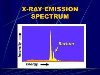
It is called characteristic because it is characteristic of the target element in the energy of the photon produced
Only K-characteristic x-rays of tungsten are useful for imaging
Bremsstrahlung Radiation • Heat & Characteristic produces EM energy by e- interacting with tungsten atoms e- of the target material • Bremsstrahlung is produced by e- interacting with the nucleus of a target tungsten atom
Bremsstrahlung Radiation • A projectile e- that completely avoids the orbital e- as it passes through a target atom may pass close enough to the nucleus of the atom to convert some of the projectile e- kinetic energy to EM energy • Because of the electrostatic force?
Bremsstrahlung is a german word meaning slowed-down radiation
X-ray energy • Characteristic x-rays have very specific energies. K-characteristic x-rays require a tube potential of a least 70 kVp • Bremsstrahlung x-rays that are produced can have any energy level up to the set kVp value. Brems can be produced at any projectile e- value
Discrete spectrum • Contains only specific values
Continuous Spectrum • Contains all possible values
Characteristic X-ray Spectrum • Characteristic has discrete energies based on the e- binding energies of tungsten • Characteristic x-ray photons can have 1 of 15 different energies and no others
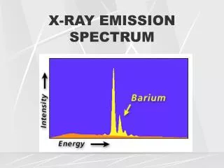
Bremsstrahlung X-ray Spectrum • Brems x-rays have a range of energies and form a continuous emission spectrum
Factors Affecting the x-ray emission spectrum • Tube current, Tube voltage, Added filtration, Target material, Voltage waveform • The general shape of an emission spectrum is always the same, but the position along the energy axis can change
Quality • The farther to the right the higher the effective energy or quality
Quantity • The more values in the curve, the higher the x-ray intensity or quantity
mAs • A change in mA or s or both results in the amplitude change of the x-ray emission spectrum at all energies • The shape of the curve will remain the same
kVp • A change in voltage peak affects both the amplitude and the position of the x-ray emission spectrum
Filtration • Adding filtration is called hardening the x-ray beam because of the increase in average energy • Characteristic spectrum is not affected & the maximum energy of x-ray emission is not affected
Filtration • Adding filtration to the useful beam reduces the x-ray beam intensity while increasing the average energy • Added filtration is an increase in the average energy of the x-ray beam (higher quality) with a reduction in x-ray quantity • Lowering the amplitude and shifting to the right
Target Material • The atomic number of the target affects both the quantity and quality of x-rays • Increasing the target atomic number increases the efficiency of x-ray production and the energy of characteristic and bremsstrahlung x-rays
Voltage Waveform • 5 voltage waveforms: half-wave rectification, full-wave rectification, 3-phase/6-pulse, 3-phase/12-pulse, and high-frequency. • Maintaining high voltage potential
X-ray Quantity or Intensity • What units of measurement is used for radiation exposure or exposure in air? • Milliampere-seconds (mAs) – x-ray quantity is proportional to mAs • Kilovolt Peak (kVp) – If kVp were doubled the x-ray intensity would increase by a factor of four or kVp2
X-ray Quantity or Intensity • Distance – x-ray intensity varies inversely with the square of the distance from the x-ray target • When SID is increased, mAs must be increased by SID2to maintain constant OD
Filtration • 1 to 3 mm of aluminum (Al) added to the primary beam to reduce the number of low-energy x-rays that reach the patient, reducing patient dose • Filtration reduces the quantity of x-rays in the low-energy range
X-ray Quality or Penetrability • As the energy of an x-ray beam is increased, the penetrability is also increased • High-energy photons are able to penetrate tissue farther than low-energy photons • High-quality = high-penetrability • Low-quality = low-penetrability
HVL = Half-Value Layer • What is the HVL • HVL is affected by the kVp and added filtration in the useful beam • Photon quality is also influenced by kVp & filtration • HVL is affected by kVp
HVL • In radiography, the quality of the x-rays is measured by the HVL • The HVL is a characteristic of the useful x-ray beam • A diagnostic x-ray beam usually has an HVL of 3 to 5 mm Al
HVL • 3 to 5 mm Al = to 3 to 6 cm of soft tissue • HVL is determined experimentally and a design specification of the equipment
X-ray Quality • Kilovolt Peak (kVp) = increasing the kVp increased photon quality and the HVL
Types of Filtration • Diagnostic x-ray beams have two filtration components – inherent filtration and added filtration • Inherent filtration – The glass enclosure of the tube (the window) – approximately 0.5 mm Al equivalent
Added Filtration • 1 or 2 mm sheet of aluminum between the tube housing and the collimator • The collimator contributes an additional 1mm Al equivalent added filtration