Biotinylation Assessment of Proteins in Cultured Hippocampal Neurons
In this experiment, primary hippocampal neurons from E18 Sprague Dawley embryos were cultured for 10 days and subjected to biotinylation. Following protein harvest and centrifugation to eliminate debris, specific proteins were incubated with NHS-LC-biotin. Subsequent separation via 10% SDS-PAGE allowed for the examination of biotinylated protein profiles. The analysis included silver staining and HRP-streptavidin detection, revealing a range of molecular weights, confirming the biotinylation of proteins beyond those visible in silver staining. This method shows the extent of biotin labeling in neuronal protein samples.

Biotinylation Assessment of Proteins in Cultured Hippocampal Neurons
E N D
Presentation Transcript
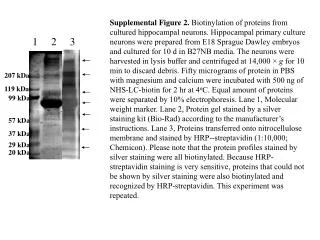
Supplemental Figure 2. Biotinylation of proteins from cultured hippocampal neurons. Hippocampal primary culture neurons were prepared from E18 Sprague Dawley embryos and cultured for 10 d in B27NB media. The neurons were harvested in lysis buffer and centrifuged at 14,000 × g for 10 min to discard debris. Fifty micrograms of protein in PBS with magnesium and calcium were incubated with 500 ng of NHS-LC-biotin for 2 hr at 4oC. Equal amount of proteins were separated by 10% electrophoresis. Lane 1, Molecular weight marker. Lane 2, Protein gel stained by a silver staining kit (Bio-Rad) according to the manufacturer’s instructions. Lane 3, Proteins transferred onto nitrocellulose membrane and stained by HRP--streptavidin (1:10,000; Chemicon). Please note that the protein profiles stained by silver staining were all biotinylated. Because HRP-streptavidin staining is very sensitive, proteins that could not be shown by silver staining were also biotinylated and recognized by HRP-streptavidin. This experiment was repeated. 1 2 3 207 kDa 119 kDa 99 kDa 57 kDa 37 kDa 29 kDa 20 kDa