Download
1 / 24
270 likes | 516 Vues
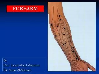
HAND & WRIST. Dr. Saeed Vohra. Dr. Jamila El- Medany. OBJECTIVES. At the end of the lecture, students should be able to: Describe the anatomy of the deep fascia of the wrist & hand (flexor & extensor retinaculae & palmar aponeurosis ).

E N D
HAND & WRIST Dr. Saeed Vohra Dr. Jamila El-Medany
OBJECTIVES • At the end of the lecture, students should be able to: • Describe the anatomy of the deep fascia of the wrist & hand (flexor & extensor retinaculae & palmar aponeurosis). • List the structures passing superficial & deep to flexor retinaculum. • Describe the anatomy of the insertion of long flexor & extensor tendons. • Describe the anatomy of the small muscles of the hand (origin, insertion action & nerve supply)
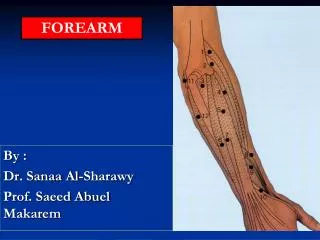
Retinacula • Flexor & Extensor Retinaculua: • Bands of Deep Fascia in front & back of Wrist • Function: • They Hold the long flexor and extensor tendons at the wrist in position. • Attachments: • Medially: Both retinacula attached toPisiform & Hook of Hamate. • Laterally: • Flexor Retinaculum attached to Tubercle of Scaphoid & Trapezium. • Extensor Retinaculum attached to Distal end of Radius
Structures Superficial to Flexor Retinaculum From Medial to Lateral Tendon of Flexor carpiulnaris. Ulnar nerve. Ulnar artery. Palmarcutaneous branch of ulnar nerve. Tendon of Palmaris longus. Palmarcutaneous branch of median nerve.
Carpal Tunnel • Formed fromConcave anterior surface of the Carpus covered by Flexor Retinaculum • Contents • From Medial to Lateral • Tendons of flexor digitorumsuperficialis & profundus • Median nerve • Flexor PollicisLongus • (Flexor carpi radialis)
Carpal Tunnel Syndrome • Causes : • Compression of the median nerve within the carpal tunnel • Manifestations: • 1. Burning pain (pins and needles ) in the lateral three and half fingers. • No paresthesia over the thenar eminence?
Carpal Tunnel Syndrome N • 2. Weakness or atrophy of the thenar muscles (Ape Hand). • Inability to Opposethe thumb.
PalmarAponeurosis • The Thickened deep fascia of the Palm. • It is Triangular in shape , occupies the central area of the palm. • Apex: • Attached to the distal border of flexor retinaculum and receives the insertion of palmarislongus. • Base: • Divides at the bases of the fingers into four slips that pass into the fingers. • Functions: • 1. Firmly attached to the overlying skin and improves the grip. • 2. Protects the underlying tendons, vessels & nerves. • 3. Gives origin to palmarisbrevis muscle.
Insertion of Flexor Dig Superficialis & Profundus • Flexor dig superficialis • Each tendon: • Divides into two halves & pass around the Profundus Tendon. • The two halves Meet on the posterior aspect of Profundus tendon (partial decussation of fibers). • Reunionof the two halves. • Further Division into two slips attached to the Bordersof Middle Phalanx. • Flexor dig Profundus • Inserted into the Base of the Distal Phalanx.
Fibrous Flexor (Digital) Sheath • A Strong Fibrous Sheath, which covers the anterior surface of the fingers and attached to the sides of the phalanges. • Its Proximal end is opened • Its Distal endis closed • The Sheath with the anterior surfaces of the phalanges & the interphalangeal joints form an Osteofibrous blind Tunnel for the long flexor tendons of the fingers.
Synovial Flexor Sheaths • Common Synovial sheath(Ulnar Bursa) • Contains tendons of Flexor DigitorumSuperficialis & Profundus • The Medialpart of the sheathextends distally (without interruption) on the tendons of the little finger. • The Lateral part of the sheath stops on the middle of the palm. • The distal ends of the long flexor tendons to(Index, Middle & Ring) fingers acquire Digital Synovila Sheaths.
Synovial Flexor Sheaths • Flexor PollicisLongustendon has its own synovial sheath (Radial Bursa) • Function of Synovial Sheaths: • They allow the long tendons to move smoothly with a minimum of friction beneath the flexor retinaculum and the fibrous flexor sheaths.
Lumbrical Muscles (4) Action: Flex metacarpophalangeal joints and extend interphalangeal joints of fingers Except thumb
PalmarInterossei(4) 2 3 4 1 2 3 4 Action: Adduction of fingers toward center of the 3rd one.
Dorsal Interossei(4) AB AB 3 2 4 1 Action: Abduction of fingers away from the 3rd one.
Action of Lumbricals & Interossei Writing position
Extensor Expansion • Formed from the expansion of the tendons of extensor dig. at the PIJ, • The tendon splits into three parts: • One Central: inserted into the base of Middle phalanx. • Two laterals: inserted into the base of the Distal phalanx. • The Expansion Receives the insertions of: • Corresponding Interosseousmuscle (on each side). • Lumbricalmuscle (on the lateral side).