Download
1 / 24
240 likes | 297 Vues
Learn about the intricacies of fertilization and implantation of the human embryo, including phases, results, and zygote formation. Understand the chromosomal uniqueness, sex determination, and cleavage process involved in early stages of development.

E N D
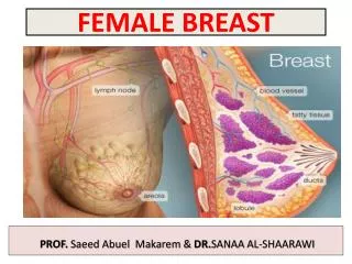
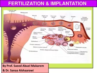
FERTILIZATION & IMPLANTATION By Prof. SaeedAbuelMakarem & Dr. SanaaAlshaarawi
OBJECTIVES • By the end of the lecture, you should be able to: • Identifyfertilizationand its site. • List the phases of fertilization. • Describe theresultsof fertilization. • Describe the formation of blastocyst. • Identify implantationand its site. • Describe the mechanism of implantation. • Describe the formation of primary chorionic villi. • List the sites of ectopic pregnancy.
FERTILIZATION Definition: It is the process during which a male gamete (sperm) unites with a female gamete (oocyte) to form a single cell (ZYGOTE).
Fertilization • It is a complex process. • It begins with a contact between sperm & ovum. • Ends up with interminglingof the maternal and paternal chromosomes.
Site of Fertilization • Usually in the ampulla of uterine tube. • Ampulla is the longest and widest part of the tube. • Fertilizationmay occur in any other part of tube. • Never occurs in the uterine cavity. • Chemical signal from oocyte attracts the sperms.
Phases of Fertilization 1&2- Passage of the sperm through the cells of the corona radiata by the effect of: a) Hyaluronidase enzymesecreted from the acrosome of the sperm. b) By movement of its tail. 3- Penetration of the zona pellucida byacrosine E. (a substance secreted from acrosomal cap). 4- Fusion of the plasma membranes of the oocyte and the sperm. 5- Completion of the second meiotic division of the oocyte & formation of the female pronucleus. 6- Formation of the male pronucleus.
CHROMOSOMES IN THE ZYGOTE • Zygoteis genetically unique. • Half of its chromosomes comes from the father and the other half comes from the mother. • New combination is formed which is different from either of the parents. • This mechanism forms biparental inheritance and leads to variation of the human species.
Sex of the Embryo • Embryo's chromosomal sex is determined at the time of fertilization by genetic studies. • Sexis determined by the type of sperm (X or Y) that fertilizes the oocyte. • So, it is the father whose gamete decides the sex. • Zonal reaction :it is a change in properties of zona pellucida that makes it impermeable to other sperms.
Results of Fertilization • Stimulates the penetrated oocyte to complete its 2nd meiotic division. • Restores the normal diploid number of chromosomes. • Determines the sex of the embryo. • Initiates cleavage (cell division) of the zygote.
Cleavage of Zygote • It is the repeated mitotic divisions of the zygote. • Normally occurs in the uterine tube. • Rapid increase in the number of the cells. • These smaller embryonic cells are now called, Blastomeres. 16-32 blastomeres
Cleavage of Zygote • It begins about 30hours after fertilization. • Zygote divides into 2, then 4, then 8, then 16 cells. • Zygote lies within the thick zona pellucida during cleavage. • Zygote migrates in the uterine tube during cleavage from lateral to medial. • Under the microscope, the zona pellucida is a translucent membrane 16-32 blastomeres
Morula • When there are 16-32 blastomeres the developing human is called MORULA. • The Morula reaches the uterine cavity at this stage. • Spherical Morula is formed about 3 days after fertilization. • It resembles mulberry or blackberry.
Mechanism of Blastocyst Formation : • TheMorula reaches theuterine cavity by the 4th day after fertilization, & remains free for one or two days. Fluid passes from uterine cavity to the Morula. • Now the Morula is called Blastocyst, its cavity is called blastocystic cavity, its cells divided into Embryoblast& Trophoblast.
BLASTOCYST A cavity appears within the morula dividing its cells into 2 groups: Outer cell layer called trophoblast. Inner cell layer (mass) calledEmbryoblastattached to one of the poles of the blastocyst. The cavity is called blastocystic cavity or blastocele.
IMPLANTATION • Definition : • It is the process by which theBlastocyst penetrates the superficial (Compact) layer of the endometrium of the uterus. • Site: • The normal site of implantationis the posterior wall of the body of the uterus near the fundus. • Time: • It begins about the 6th day after fertilization. • It is completedby the 11th or12th day.
Mechanism of Implantation • Bythe 5th day the Zona pellucida degenerates. • Blastocystbegins implantation by the 6th day. • Trophoblast cells at the embryonic pole of the balstocyst begine to penetrate the epithelium of the endometrium (uterine mucosa) at the 6th day of development. • Penetration results from proteolytic enzymes (eg.COX-2) produced by the trophoblast.
By 6th day the blastocystadheres to the endometrium (A) and beginning of penetration. • By 7th day, Trophoblast differentiated into 2 layers: (B) Cytotrophblast, inner layer, mitotically active. Syncytiotrophoblast(outer multinucleated mass, with indistinct cell boundary) ; Invasion of endometrium continues with the syncytiotrophoblasts. • By 8th day the blastocyst issuperficially embedded in the compact layer of the endometrium. B
Blood-filled Lacunae appear in the Syncytiotrophoblast which communicate forming a lacunar network by the 10th or 11th day. • Syncytiotrophoblast erodes the endothelial lining of thematernal capillarieswhich known as sinusoids. Now blood of maternal capillaries reaches the lacunae so, Uteroplacental circulation is established by 11th or 12thday.
Endometrial cells undergo a process calledapoptosis (programmed cell death) to facilitates invasion of endometrium by the Syncytiotrophoblast. Syncytiotrophoblastengulf these degenerated cells for nutrition of the embryo. Implantation can be detected by: 1-Ultrasonography. 2-Pregnancy test (hCG ) (Home Pregnancy Test): (human chorionic gonadotrophin) hormone is secreted by the Syncytiotrophoblast about the endof 2nd week. (HCG can be measured in both the blood and urine to determine if a woman is pregnant).
Early Pregnancy Factor(EPF) • Is an immunosuppressant protein; • Its functionis to prevent the immune system from attacking the new embryo. • Secretedby trophoblast cells. • Appears inmaternal serum within 24--48 hrs., after fertilization. • It is the basis for EPT (Early pregnancy test)in the first 10 days of development.
Formation of The Primary Chorionic villi • By the 13th day Proliferationof Cytotrophblast cells produce extension inside the Syncytiotrophoblastto form the primary chorionic villi.
Ectopic Implantation (Pregnancy) • The usual site of implantation is the posterior wall of the body of uterus (X). • Tubal pregnancy is the most common type of ectopic pregnancy (A). • Ovarian pregnancy is the least common typeof ectopic pregnancy (H).
Ectopic Pregnancy Ectopic Pregnancy: 1- Placenta Previa. 2- Tubal. 3- Ovarian. 4- Abdominal. 5- Pelvic. 6- Cervical. Placenta previa centralis • It means implantation outside the uterine cavity. 1. 95 to 97% of ectopic pregnancies occurs in the uterine tube. • Most are in the ampulla & isthmus. 2. Placenta previa: • Implantation occurs in the loweruterine segment. Placenta previa lateralis Placenta previa marginalis