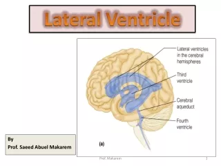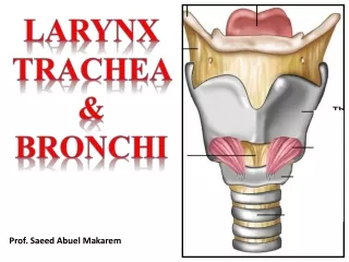
By Prof. Saeed Abuel Makarem
210 likes | 907 Vues
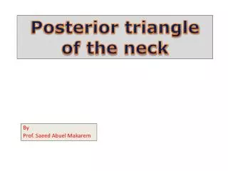
Posterior triangle of the neck. By Prof. Saeed Abuel Makarem. STERNOCLEIDOMASTOID. The sternocleidomastoid or sternomastoid is a strap- like muscle that descends obliquely across the side of the neck. . TRIANGLES OF THE NECK.

By Prof. Saeed Abuel Makarem
E N D
Presentation Transcript
Posterior triangle of the neck By Prof. Saeed Abuel Makarem
STERNOCLEIDOMASTOID • The sternocleidomastoid or sternomastoid is a strap- like muscle that descends obliquely across the side of the neck.
TRIANGLES OF THE NECK The neck is divided into anterior and posterior triangles by the sternocleidomastoid muscle; • Anterior triangle lies in front of the muscle • and • Posterior triangle lies behind the muscle.
Boundaries: Anteriorly: Posterior border of sternomastoid. Posteriorly: Anterior border of Trapezius. Base: Middle 1/3 of the clavicle. Apex: meeting of Trapezius & Sternomastoid.
OMOHYOID MUSCLE Omohyoid muscle has: • Inferior belly, • Intermediate tendon, • and • Superior belly.
The inferior belly of omohyoid subdivides the posterior triangle into: • a large occipital triangle above and • a small supraclavicular triangle below.
Roof: Skin, Superficial fascia, whichcontains:Platysma muscle Cutaneous branches of cervical plexus External jugular vein& Investing layer of deep cervical fascia. Floor: from below upward: • Scalenus medius, • Levator scapulae, • Splenius capitis, and • Semispinalis capitis. The muscles of the floor are covered by prevertebral layer of deep cervical fascia. NB. Scalenus anterior is hidden by the sternomastoid muscle
The deep fascia of the neck is called deep cervical fascia • Parts of Deep Cervical Fascia: • Investing layer. • Prevertebral layer. • Pretracheal layer. • Carotid sheath.
PLATYSMA • The platysma can be seen as a sheet of muscle by asking the patient to clench his jaw firmly. • The muscle extends from the body of the mandible downward over the clavicle onto the anterior thoracic wall.
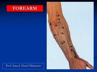
Contents of Posterior triangle Nerves: The main contents are nerves: 1- The three trunks of the brachial plexus. 2- Four cutaneous branches of cervical plexus. (lesser occipital, great auricular, transverse cervical & supraclavicular nerves. 3- Spinal accessory. Arteries: 1- 3rd part of subclavian artery. 2- Suprascapular artery. 3- Transverse cervical artery. 4- Occipital artery. Veins: 1- Subclavian vein. 2- External jugular vein. Muscle: Inferior belly of omohyoid muscle.
ACCESSORY NERVE (SPINAL PART)11th cranial nerve • The spinal part of the accessory nerve enters the posterior triangle by emerging from beneath the middle of the posterior border of sternomastoid muscle, which it has supplied. • It runs downward and laterally across the posterior triangle on the levator scapulae muscle. • It also supplies the trapezius.
EXTERNAL JUGULAR VEIN The external jugular vein begins just behind the angle of the mandible by the union of the posterior auricular vein & the posterior division of the retromandibular vein.
The external jugular vein descends obliquely superficial to sternomastoid muscle and, just above the clavicle in the posterior triangle, pierces the deep fascia and drains into the subclavian vein. • It is the only tributary of the subclavian vein. • It varies considerably in size, and its course extends from the angle of the mandible to the middle of the clavicle.
THIRD PART OF SUBCLAVIAN ARTERY The third part of the subclavian artery occupies the lower anterior angle of the posterior triangle just behind the sternomastoid muscle.